Yichi (Tony) Zhang, PhD
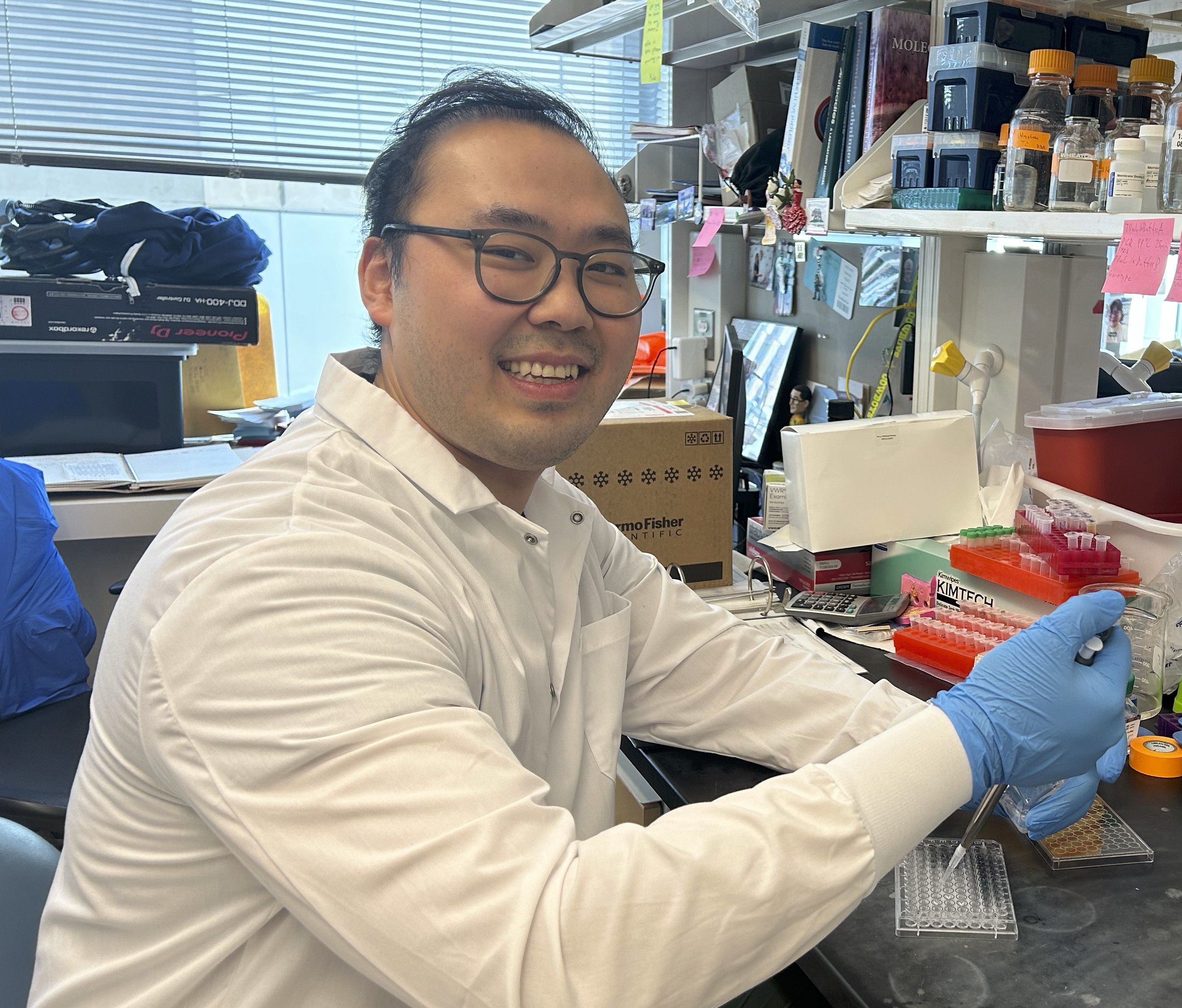
Cancer cachexia, characterized by progressive muscle wasting and weight loss in cancer patients, is a common and multifaceted syndrome that negatively impacts patient quality of life. Cachexia has no available treatments to date, due to insufficient knowledge about the underlying mechanisms. Cachexia is particularly prominent in pancreatic cancer patients. Previous work from the Vander Heiden lab has identified that there is diminished secretion of pancreatic enzymes due to the pancreatic cancer, which contributes to tissue wasting because breakdown of muscle tissue can release amino acids into circulation. Dr. Zhang is interested in understanding whether amino acids released from muscle are fueling tumor growth or sustaining the host organism. Specifically, as circulating amino acids are often used by the liver to produce glucose, Dr. Zhang wants to study how liver glucose metabolism is affected in pancreatic cancer cachexia. This work seeks to improve the quality of life of cancer patients by reducing cachexia while seeking to understand how cancer impacts the whole-body metabolism of the host. Dr. Zhang received his PhD from University of Texas Southwestern Medical Center, Dallas, his MS from Carleton University, Ottawa, and his BScH from Queen’s University, Kingston.
Xianfeng Zeng, PhD
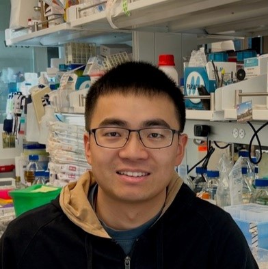
Emerging evidence underscores the profound impact of the gut microbiome, a collection of microorganisms within our digestive system, on cancer. These microorganisms collectively generate various metabolites that can significantly influence cancer progression and treatment outcomes. Dr. Zeng is employing synthetic communities and mouse cancer models to delve into the intricate connections between cancer and the microbiome. His synthetic communities, comprised of over 100 strains, allow for precise manipulation of the microbiome to elucidate the role of specific microbial metabolites in cancer. Additionally, Dr. Zeng is studying community-scale metabolism and using genetically edited strains to design synthetic communities with desired metabolic profiles. These approaches will gain valuable insights into microbiome-cancer interactions and establish a broadly applicable strategy to harness the therapeutic potential of gut microbiome. Dr. Zeng received his PhD from Princeton University, Princeton and his BS from Tsinghua University, Beijing.
Teng Gao, PhD
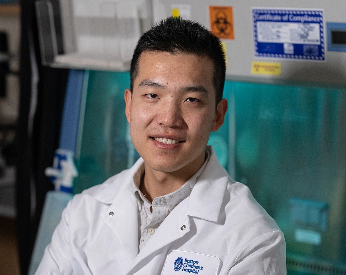
Hematopoietic stem cells, which are found in the bone marrow and give rise to all other blood cells, maintain lifelong blood production and immune function. Due to their remarkable ability to regenerate the entire blood system, medical uses of HSCs have provided cures for many previously incurable diseases, including blood cancers. However, several unanswered questions limit our ability to full harness their therapeutic potential for cancer treatment. What regulates HSC regeneration? Why does their function decline with age? How does HSC behavior vary in healthy individuals? Using cutting-edge single-cell analyses and computational biology, Dr. Gao [HHMI Fellow] aims to identify the molecular and cellular factors involved in HSC regeneration, as well as possible targets for enhancing their regenerative potential. This work could enable significant improvements in stem cell-based therapies for cancer treatment. Dr. Gao received his PhD from Harvard University, Cambridge and his BS from Washington University, St. Louis.
Tamar Kavlashvili, PhD
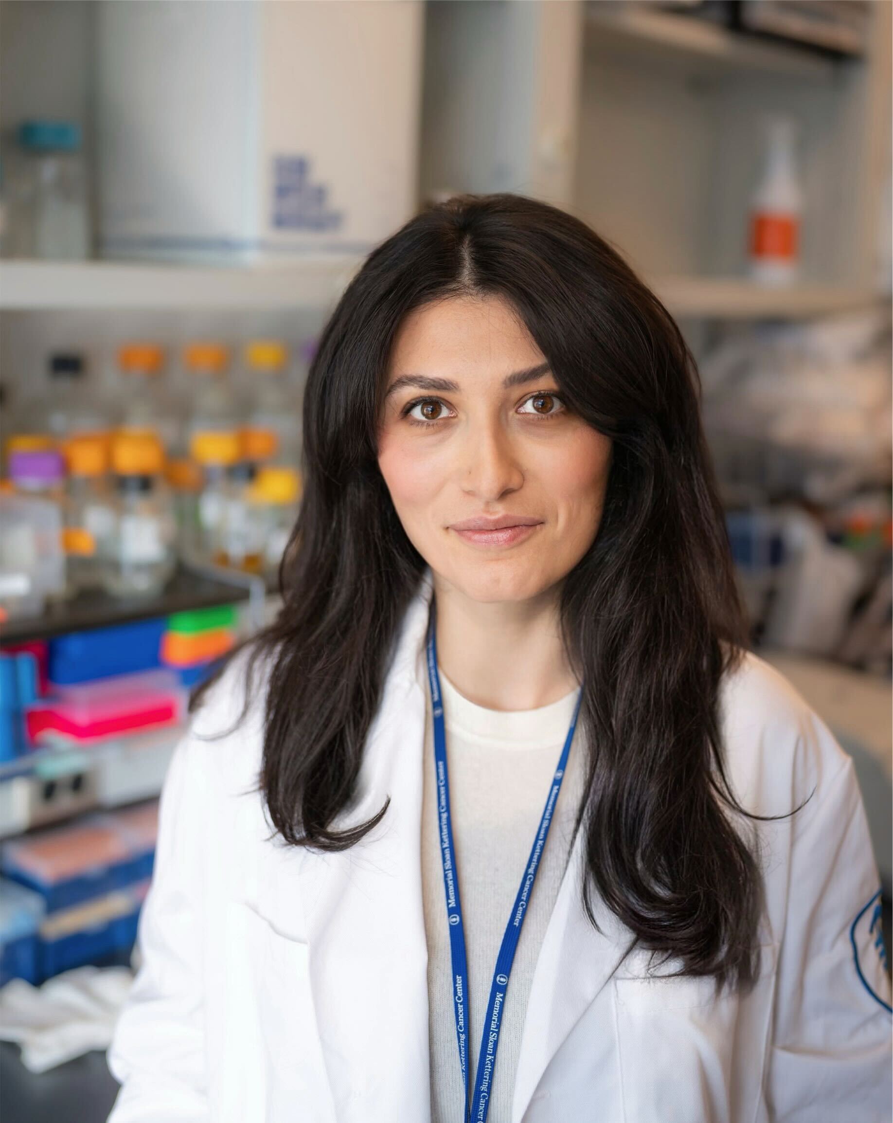
Mitochondria harbor independent genetic material known as mitochondrial DNA (mtDNA). This compact, circular molecule encodes proteins essential for the assembly of the mitochondrial electron transport chain to generate energy in form of ATP. Like nuclear DNA, mtDNA is susceptible to damage and mutations. One of the most common disease-causing aberrations of mtDNA is termed “common deletion.” This aberration disrupts mitochondrial function, resulting in neuromuscular diseases and potentially certain cancers, including colorectal cancer. Due to a lack of tools to modify the mitochondrial genome, researchers currently do not understand the mechanisms behind common deletion. Dr. Kavlashvili [Timmerman Traverse Fellow] aims to investigate by using cutting-edge molecular biology tools to edit and visualize mtDNA genomes. She will then be poised to unravel impacts of this deletion on various tissues, in order to ultimately mitigate its pathological impact. Dr. Kavlashvili received her PhD from Vanderbilt University, Nashville and her BS from University of Iowa, Iowa City.
Sue Im Sim, PhD
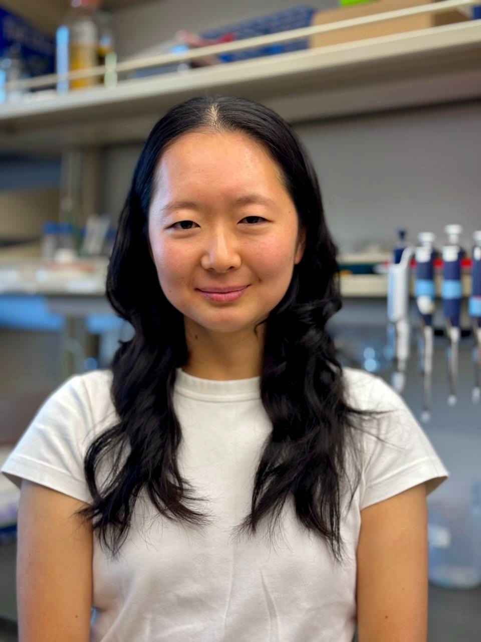
To power directional movement, cells build dynamic sheet-like protrusions at their leading edge. How individual molecules are coordinated to produce these changes in cell morphology is poorly appreciated. Dr. Sim [Connie and Bob Lurie Fellow] uses immune cell migration as a model system to investigate the self-organization of a protein assembly known as the WAVE complex, which facilitates the formation of these protrusions in migratory cells. Her work will harness recent advances in electron microscopy and protein prediction and design to study the mechanism of the WAVE complex. As a critical player in cell migration, the dysregulation of the WAVE complex is associated with tumor cell invasion and metastasis in several cancer types. This aberrant migration enables cancer cells to travel to and infiltrate adjacent tissue sites. Understanding the fundamental mechanisms of cell migration can thus better inform the development of therapeutics that limit the progression of cancer. Dr. Sim received her PhD from the University of California, Berkeley and her BA from Bowdoin College, Brunswick.
Shreoshi Sengupta, PhD

Studies have shown that lung tumors are sustained through the formation of new blood vessels from pre-existing ones in a process called angiogenesis. Moreover, tumor cells secrete signaling proteins that help them communicate with each other and evade immune detection. However, most of these studies have been on late-stage lung tumors; our understanding of cell-cell interactions in the tumor environment during lung cancer initiation and early stages remains poor. Dr. Sengupta [Deborah J. Coleman Fellow] plans to identify the gene expression patterns in tumor cells, endothelial cells (blood-vessel-forming cells), and immune cells over time to understand how they engage in this cellular crosstalk, promoting tumorigenesis. She also plans to examine cell-cell interactions in early-stage lung cancer using organoids, or artificially grown miniature organs. This line of investigation will help understand the mechanisms underlying tumor initiation and lead to novel biomarkers that can help detect lung cancers earlier. The findings will also help identify novel therapeutic targets that can be inhibited to improve patient responses and survival. Dr. Sengupta received her PhD from Indian Institute of Science, Bangalore and her MS and BS from University of Calcutta, Kolkata.
Sangin Kim, PhD
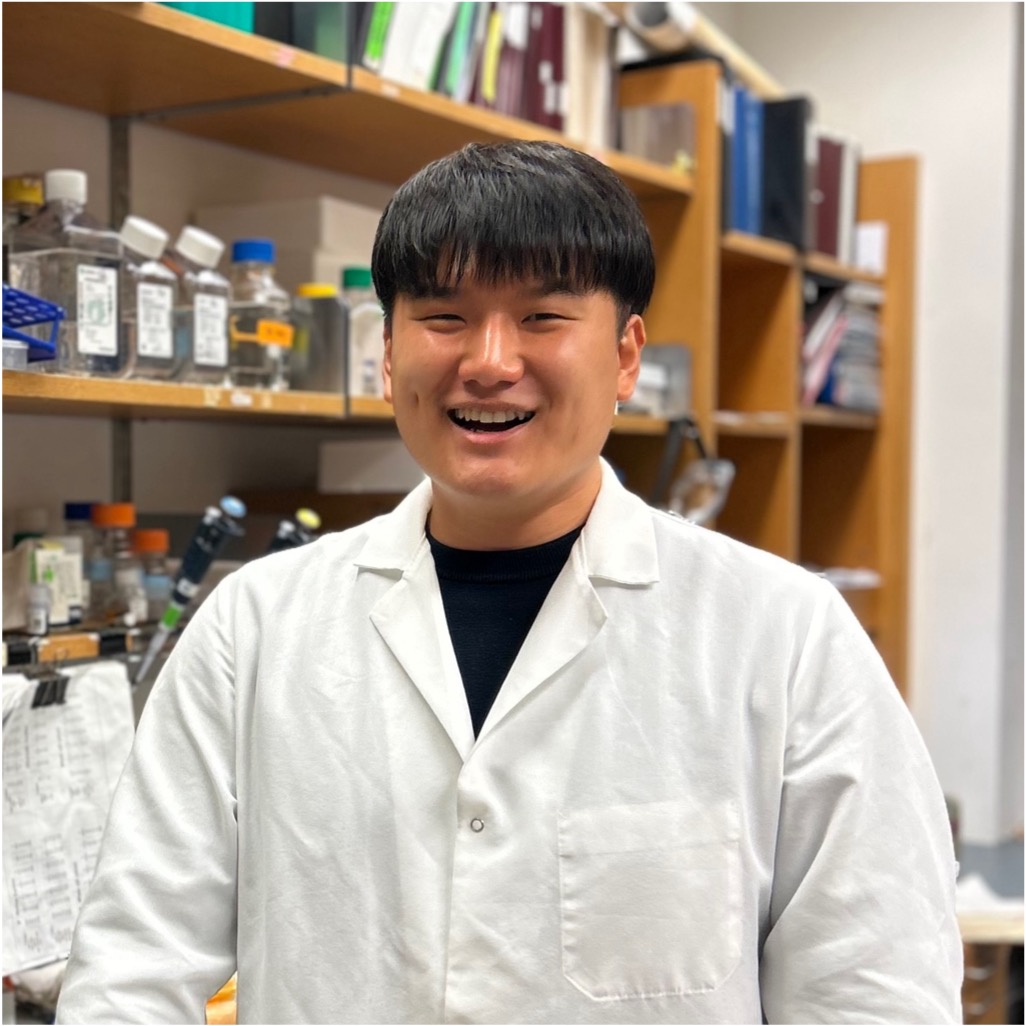
The cellular response to DNA damage is coordinated by an enzyme known as ATM kinase. Mutations in ATM are found in approximately 1% of the population and contribute to an increased risk of both hereditary and sporadic cancers, including breast cancer. Dr. Kim’s research investigates how ATM suppresses the production of double-stranded RNAs (dsRNAs) in response to DNA damage. These dsRNAs play a critical role in tumor progression. Dr. Kim aims to identify the key molecular players involved in ATM-mediated suppression of dsRNAs and elucidate how the loss of ATM function triggers inflammatory responses through dsRNA sensing pathways. By uncovering these mechanisms, Dr. Kim aims to deepen our understanding of how ATM mutations drive cancer development and uncover novel therapeutic strategies for ATM-associated cancers. Dr. Kim received his PhD and BS from the Ulsan National Institute of Science and Technology, Ulsan.
Saket Rahul Bagde, PhD
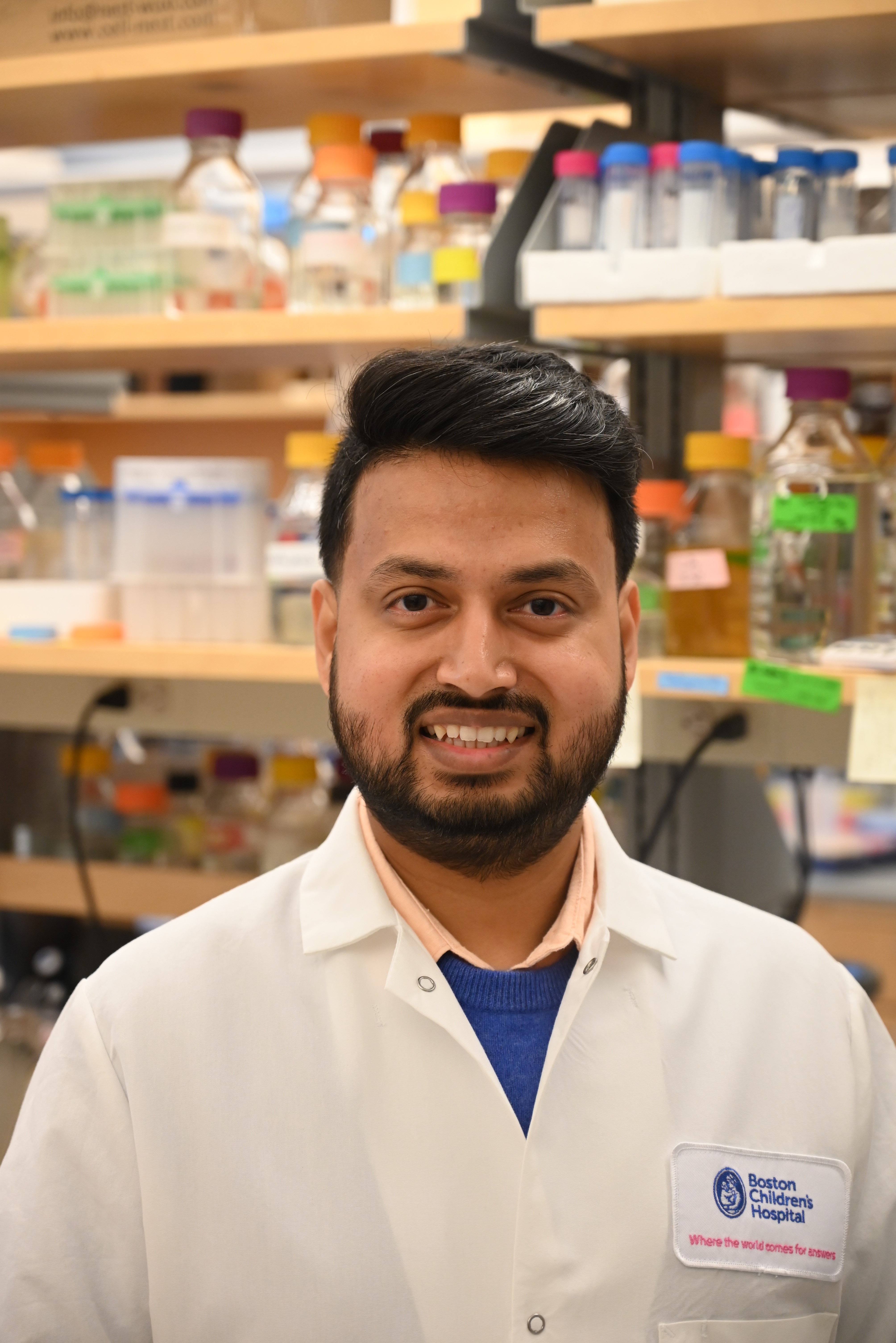
Most cancers develop in the epithelial tissue, which includes the skin and internal organ linings. Hemidesmosomes (HDs) are adhesive structures that anchor epithelial cells to the underlying base layer and maintain tissue integrity. While HD disassembly occurs normally during wound healing, tumor cells can exploit this process to detach and spread to other parts of the body. Dr. Bagde is studying how HD components interlock like Lego blocks to form stable HDs in healthy tissues and how they disassemble in cancerous tissues. To investigate this phenomenon, Dr. Bagde plans to develop organoids—self-organizing mini-organs grown in a petri dish to study disease progression. By creating simple base layers that simulate the supportive properties of the native organ base layer, he plans to promote the growth of both normal and cancerous organoids. This work has the potential to support the development of personalized cancer therapies based on patient-derived tumor samples. Dr. Bagde received his PhD from Cornell University, Ithaca and his MS and BS from the Indian Institute of Science Education and Research, Pune.
Rodrigo Gier, PhD
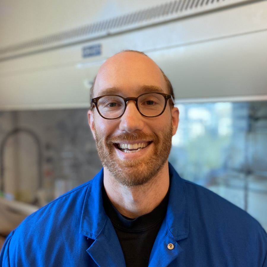
Drug therapies that selectively target proteins that drive the growth of tumor cells are rapidly becoming the standard of care for many cancers. However, tumors are often able to evade inhibition by targeted anti-cancer drugs by activating other proteins, leading to drug resistance. Dr. Gier [HHMI Fellow] is developing a new therapeutic approach that repurposes existing drugs to release highly toxic cargoes, known as payloads, that aggregate in drug-resistant cancer cells and kill them. As a general platform, it is applicable to a wide range of solid and liquid cancers. Dr. Gier received his PhD from University of Pennsylvania, Philadelphia and his BA from Swarthmore College, Swarthmore.
Pu Zhang, PhD
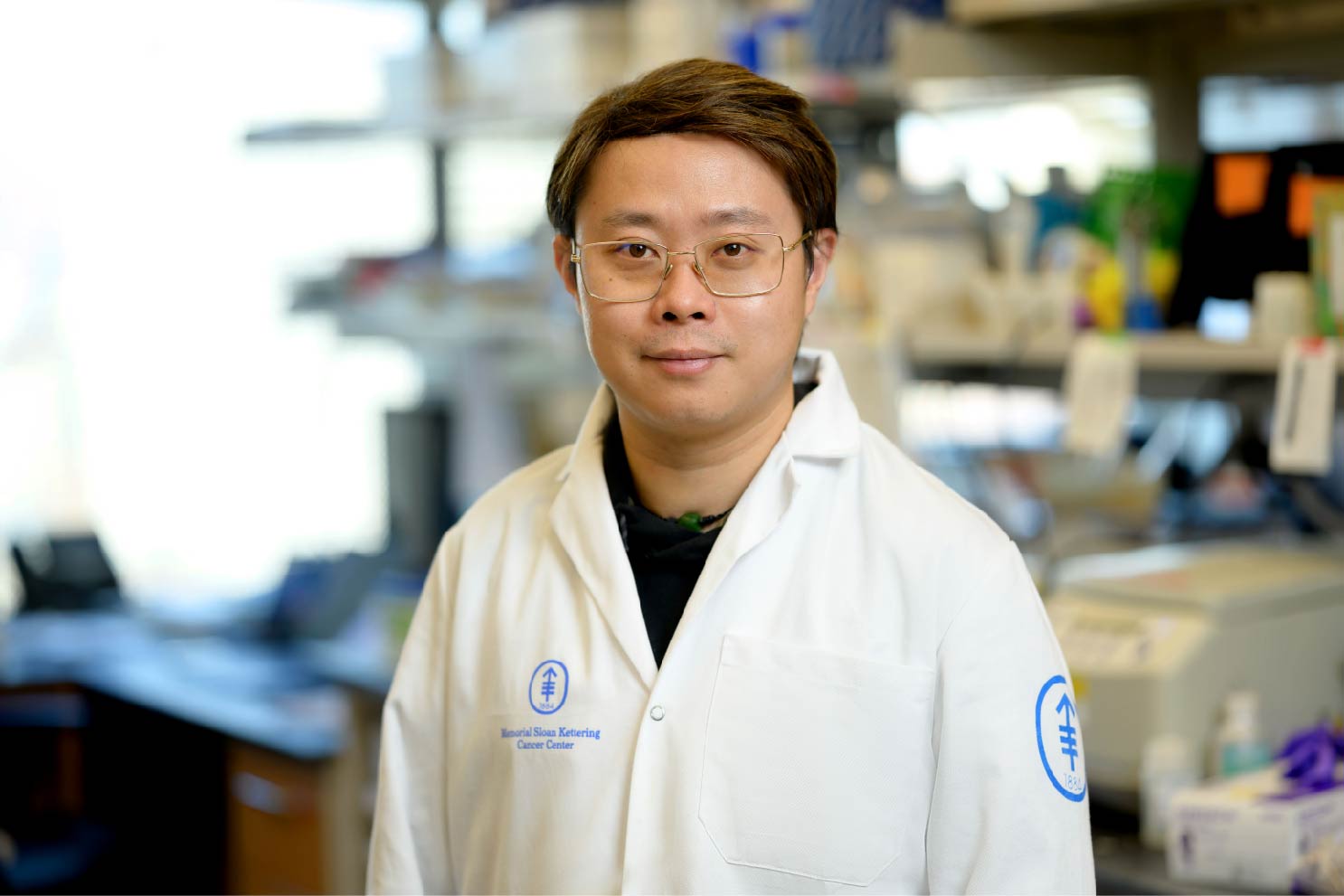
Dr. Zhang is studying a unique three-stranded nucleic acid structure, called an R-loop, to understand its role in cancer development and find ways to target and control its formation. R-loops consist of a DNA-RNA hybrid and a displaced strand of DNA. R-loops occur frequently in human genomes, and while they play an important role in blood cell differentiation and immune cell function, they can also interfere with DNA repair and promote genome instability, giving rise to leukemia. However, the dynamic nature of R-loop formation hampers the detection of this structure in a small cell sample. To address this challenge, Dr. Zhang is developing novel techniques to map R-loops in normal blood stem cells versus blood cancer cells at single-cell resolution. He also plans to investigate leukemia-specific R-loops in vitro and in vivo with CRISPR-based screening techniques. The goal of his research is to aid development of therapeutic interventions for R-loop-related gene expression dysregulation in cancer, especially leukemia. Dr. Zhang received his PhD from Ohio State University, Columbus, his MS from University of Edinburgh, Edinburgh, and his BS from Chongqing University, Chongqing.
Longyue Lily Cao, MD, PhD
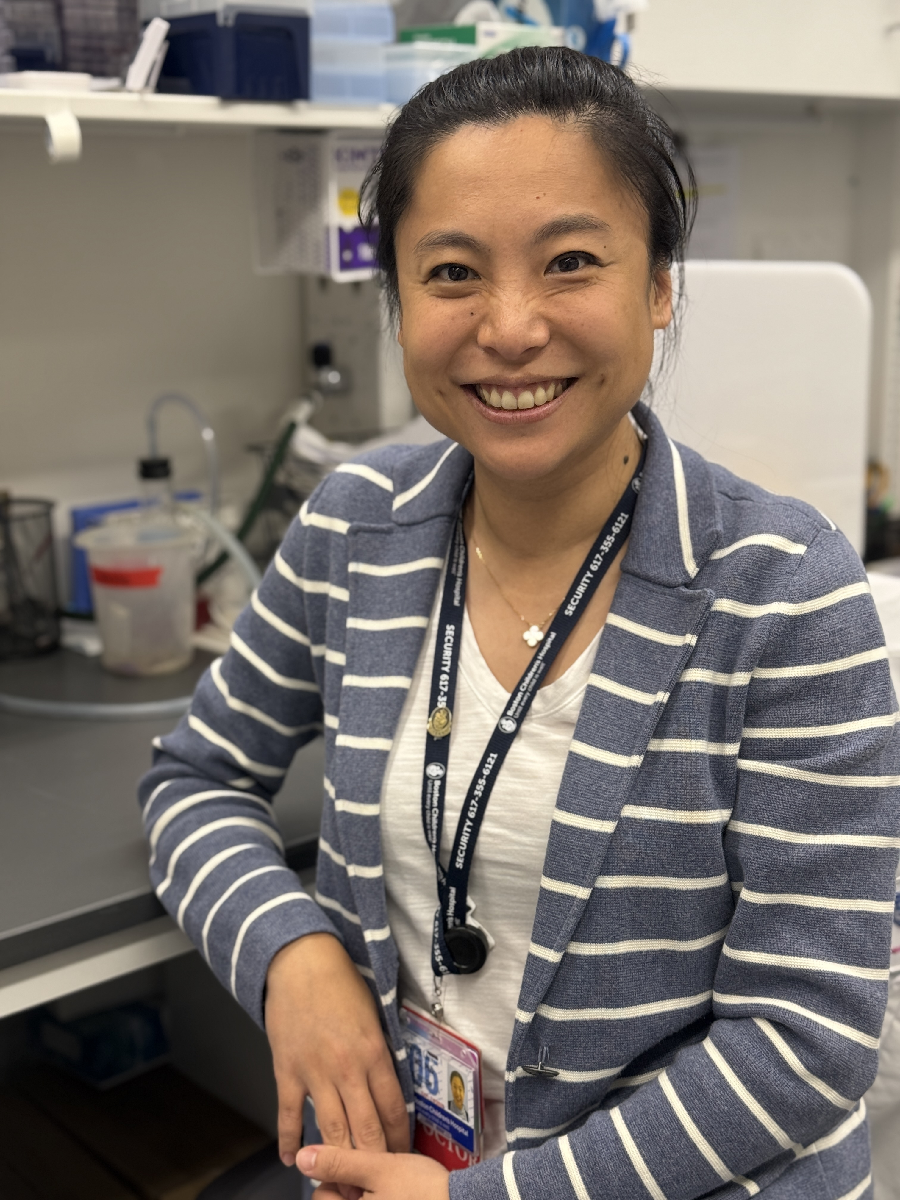
Hepatocellular carcinoma (HCC) is the most common liver cancer and has one of the highest cancer-related mortality rates. Conventional cancer immunotherapies, which largely focus on enhancing T cell activity, are unfortunately effective in only a small minority of HCC patients. Though dendritic cells (DCs) are essential for T cell activation, their potential as an immunotherapeutic target remains poorly understood. Dr. Cao is investigating how a unique, hyperactivated state of DCs can be harnessed to enhance anti-tumor immunity in a genetically engineered mouse model of HCC. Her work aims to uncover how hyperactivated DC responses generate stronger and longer-lasting protection against HCC and hopefully other cancers that are poorly responsive to conventional therapies. Dr. Cao received her MD, PhD from Albert Einstein College of Medicine, Bronx and her BS from Cornell University, Ithaca.
Laura Crowley, PhD
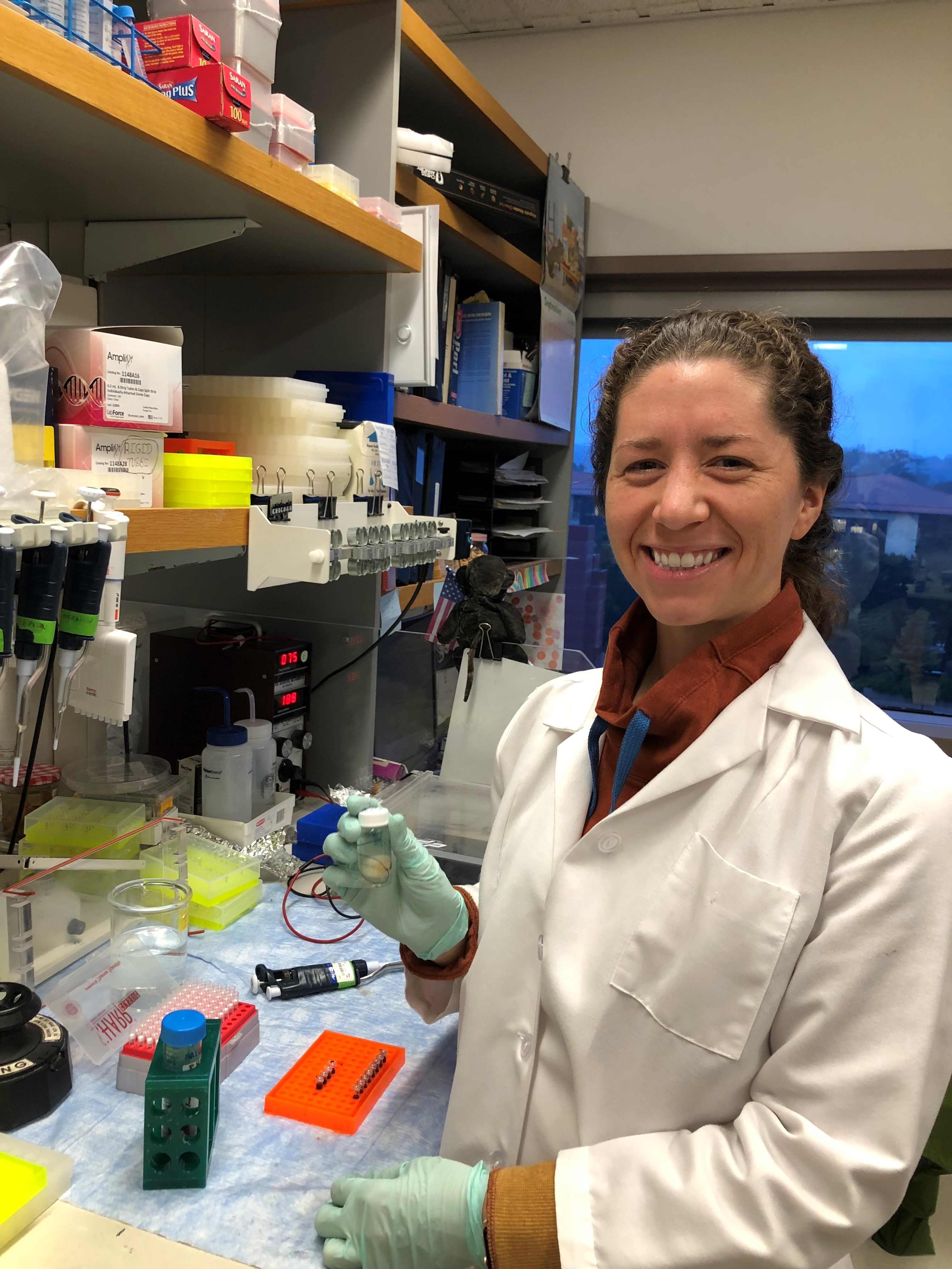
Fibroblasts are one of the earliest known cell types and they contribute to many of the most burdensome lung diseases, including cancers, fibrosis, and emphysema; however, they are surprisingly poorly understood. Dr. Crowley [HHMI Fellow] will examine the different types of fibroblasts in the mouse lung to determine where they come from and how they function normally, as well as how they change with injury and disease. This will establish an important baseline for how these cells function in mice and also provide critical, long-term insights into how these cells may function in humans, where lung diseases are very difficult to treat and are among the leading causes of mortality worldwide. Though her work will directly analyze the fibroblasts and microenvironment around lung tumors, her findings could translate to many other solid tumor contexts. Dr. Crowley received her PhD from Columbia University, New York and her BA from Colby College, Waterville.
Kheewoong Baek, PhD
Attaching a small molecule known as ubiquitin to a protein, in a process called ubiquitylation, targets that protein for degradation. By utilizing the ubiquitylation machinery, scientists are now able to target cancer-causing proteins for degradation, a strategy that has proven effective with drugs such as Lenalidomide/Revlimid to treat multiple myeloma. One way to bring proteins in proximity to ubiquitin ligases (attachers) is with synthetic adhesion molecules, or “molecular glue.” This may provide a means of targeting proteins previously deemed undruggable, including those that lack a binding site for inhibitors. Dr. Baek aims to expand the degradable proteome by establishing a formula for the design of molecular glues to target cancer-causing proteins as a therapeutic modality. Dr. Baek received his PhD from Technical University of Munich and Max Planck Institute of Biochemistry, Munich, his MS from University of Tennessee Health Science Center, Memphis, and his BA from Rutgers University, New Brunswick.
Kashish Jain, PhD
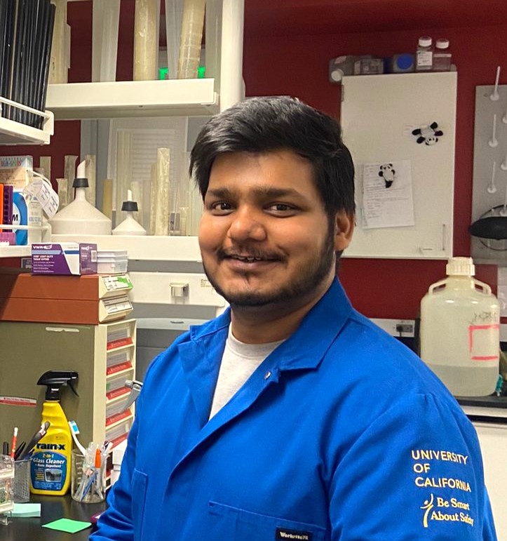
A majority of pancreatic cancer cases harbor a mutation in the KRAS gene, which is involved in cancer initiation, progression, and chemotherapy resistance. Drugs targeting KRAS mutations are often met with resistance due to limited drug penetration into the tumor. Since pancreatic cancer progression involves increased tissue stiffening, KRAS signaling might be controlled by tissue stiffness. Dr. Jain is studying the mechanisms that underlie tissue stiffness-dependent KRAS signaling at the molecular level. Understanding these mechanisms will uncover new ways to block aberrant KRAS signaling or reduce the effects of tissue stiffness on cancer progression, ultimately informing new combination therapies with KRAS-targeting drugs. Dr. Jain received his PhD from the National University of Singapore, Singapore and his BTech and MTech from Indian Institute of Technology, Kanpur.
Jordan B. Jastrab, MD, PhD
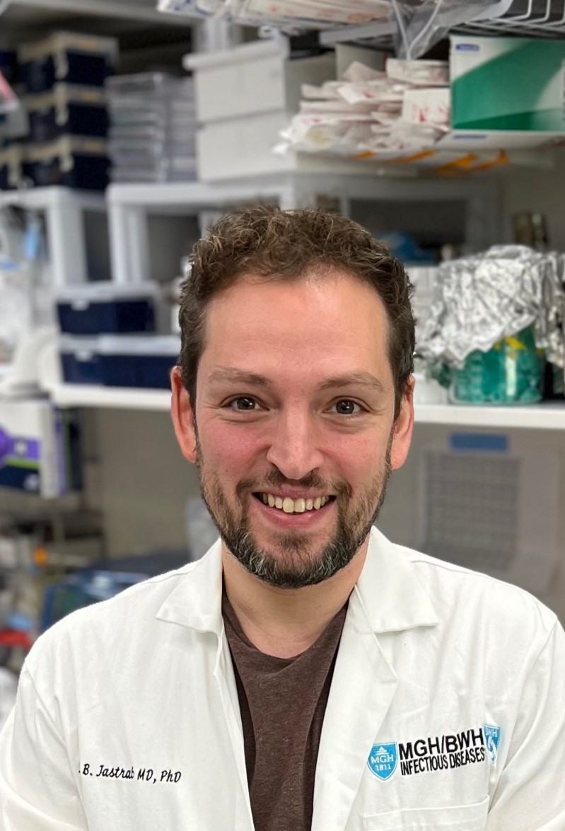
Immune cells called macrophages can swallow bacteria and contain them in membrane-bound compartments called phagosomes. From inside the phagosome, some bacteria stimulate immune pathways in the cytosol, but it is unclear how immune signals are transmitted across the membrane from the phagosome into the cytosol. To investigate, Dr. Jastrab [Robert Black Fellow] has developed a macrophage infection model using mutants of the bacterium Staphylococcus aureus that stimulate an immune complex in the cytosol called the inflammasome. He aims to identify the host and microbial pathways that facilitate activation of the inflammasome during infection. Because the activation of cytosolic immune receptors by phagosomal bacteria may be important in protection against colorectal cancer, Dr. Jastrab’s work aims to elucidate pathways that may be manipulated to prevent tumorigenesis and enhance anti-tumor immunity. Dr. Jastrab received his MD, PhD from New York University School of Medicine, New York and his BS from Tufts University, Medford.
John Devany, PhD
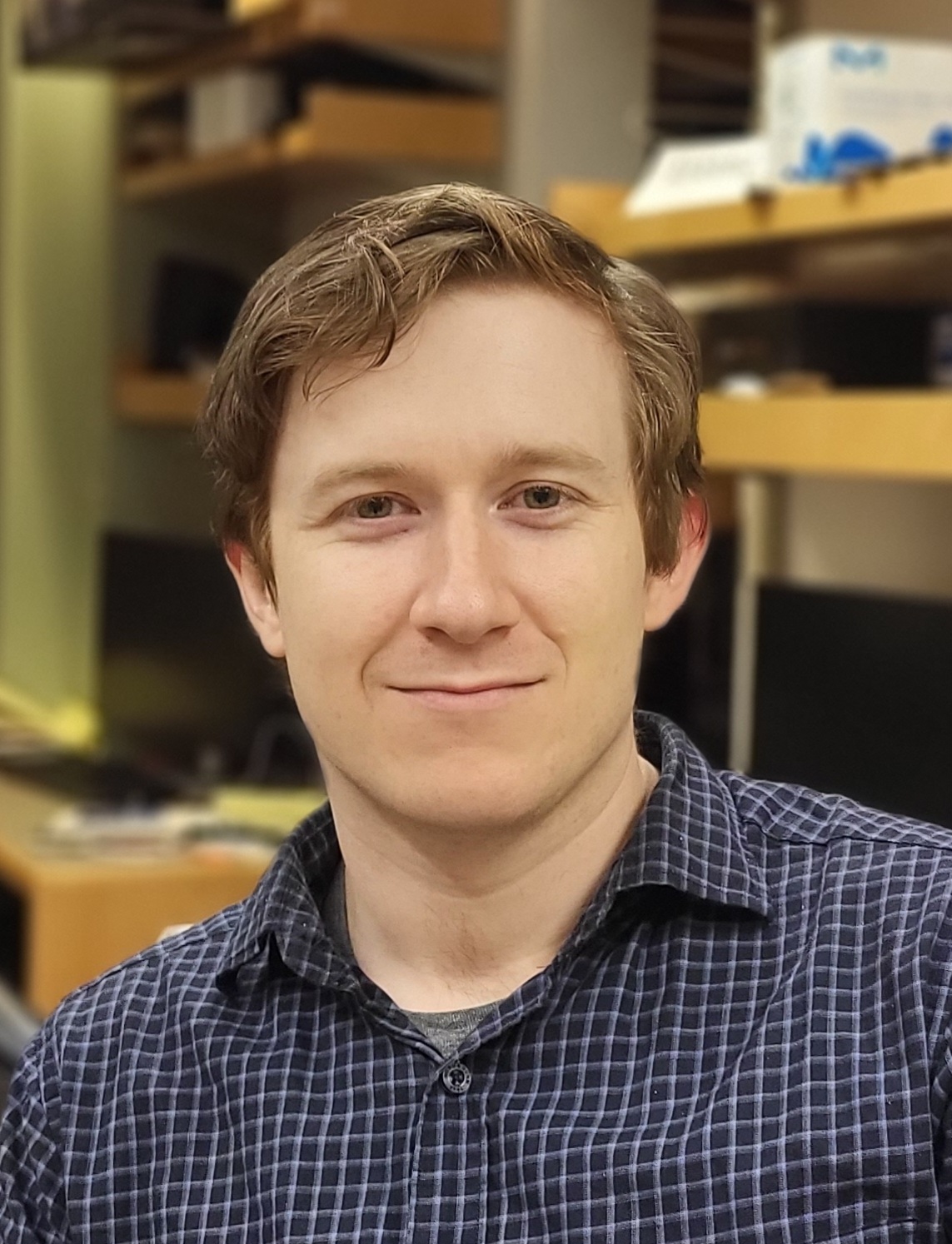
T cell therapies have led to promising results in treating blood cancers, but new approaches are required to translate these results to solid tumors. In solid tumors, T cells face unique challenges in the tumor microenvironment (TME), which limits the persistence and efficacy of adoptive T cell therapies. In T cell lymphomas (TCLs), tumor cells overcome many of the same challenges through acquired mutations. Fueled by natural selection, tumor mutations produce novel and elegant solutions to address T cell deficits in the TME. Understanding that these modifications may be superior to current bioengineering capabilities, Dr. Devany [Bakewell Foundation Fellow] plans to introduce gain-of-function mutations into therapeutic T cells to grant them the ability to survive, proliferate, and function in the TME. He will determine how each mutation restores different aspects of T cell function, allowing for the design of combinations of mutations that act synergistically. His results will aid in the development of next-generation T cell therapies to cure solid tumors. Dr. Devany received his PhD from University of Chicago, Chicago and his BS from University of California, Santa Barbara.
James C. Taggart, PhD
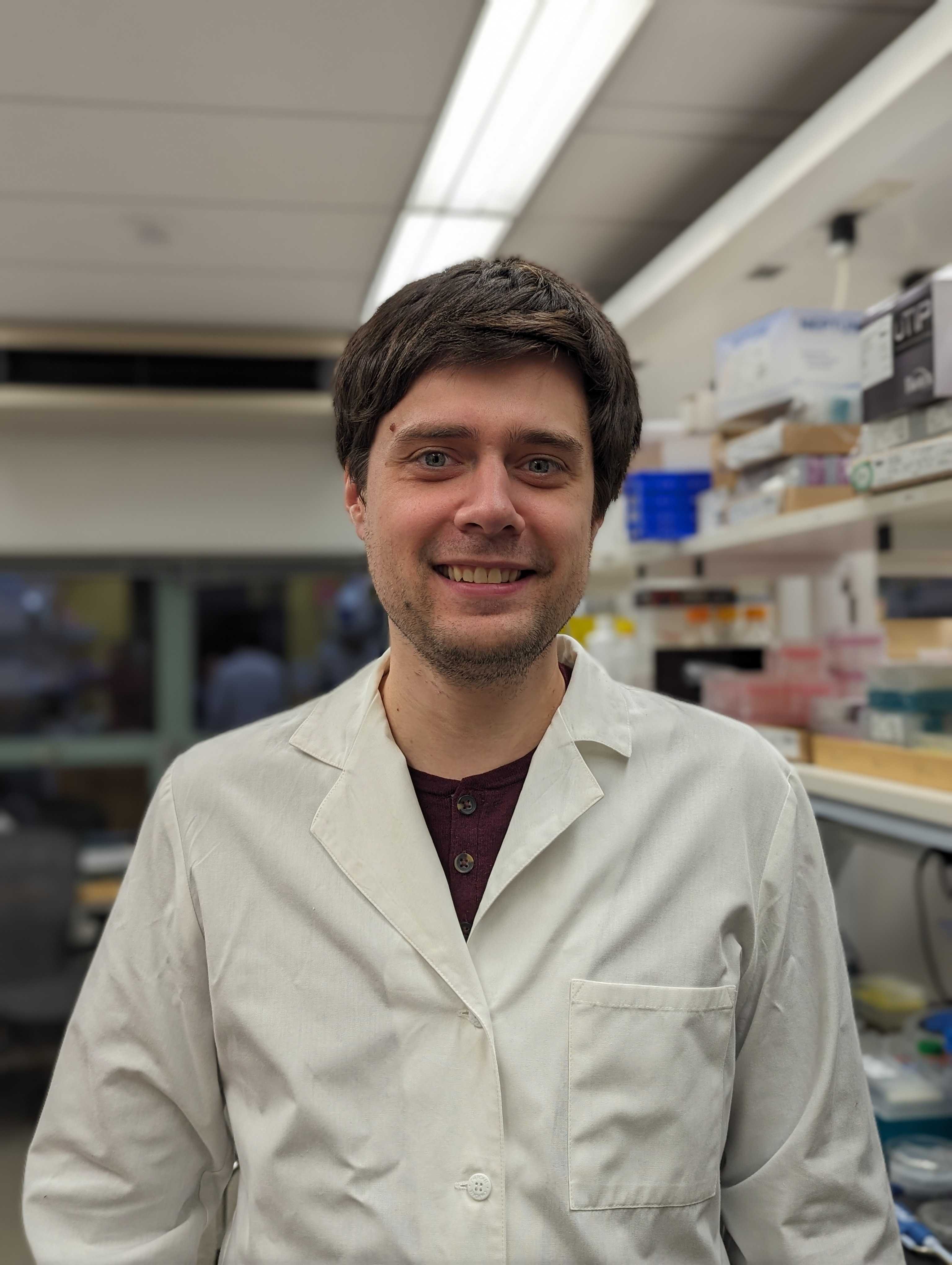
Antimicrobial resistance is a growing crisis that imperils our ability to protect patients immunocompromised by cancer treatment. Despite this, the few new antibiotics currently in clinical trials primarily use established mechanisms of action. Identification of new targets for antimicrobial drugs is thus an urgent clinical need. Recent work has shown that bacteria can tolerate substantial inhibition of many proteins thought to be essential for growth, rendering them poor drug targets. The mechanisms that cause this robustness are poorly understood. By combining cutting-edge microfluidic technologies with methods for controlled gene repression, Dr. Taggart will systematically identify mechanisms that allow bacterial cells to tolerate inhibition of genes critical for cellular growth. This work will guide the selection of targets for future antibiotic development and may reveal mechanisms by which to sensitize bacterial cells to existing drugs. Dr. Taggart received his PhD from Massachusetts Institute of Technology, Cambridge and his BS from Haverford College, Haverford.
Isabella Evavold, PhD
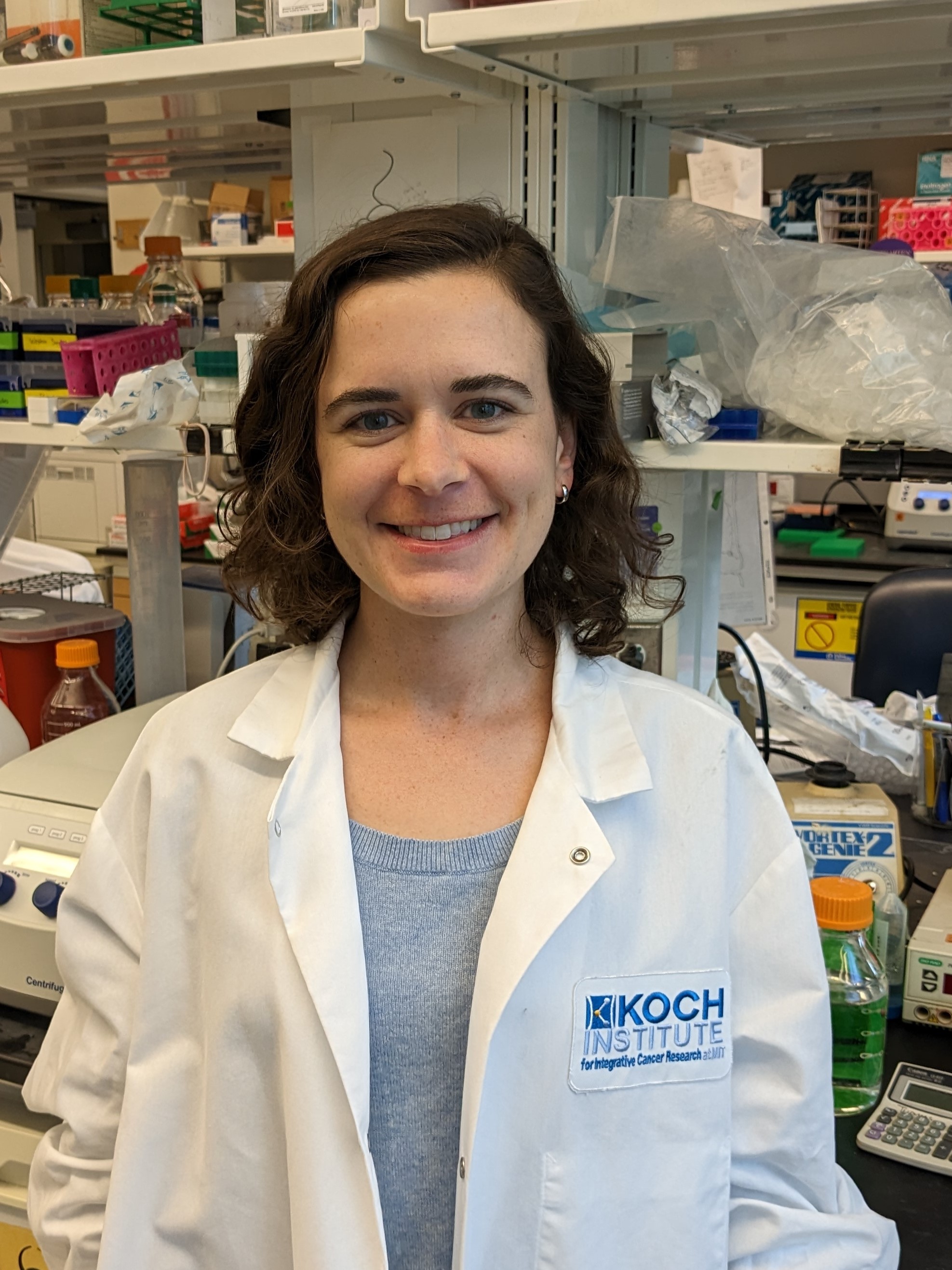
Pancreatic cancer remains unresponsive to current chemotherapy and immunotherapy treatments. However, with the recent development of mRNA vaccines and drugs that target cancer cell mutations, there is hope for a new generation of immune-based therapies. The ability of adaptive immune cells, called cytotoxic T cells, to kill cancer cells is central to anti-tumor immunity. Using mouse models of human pancreatic cancer, Dr. Evavold [Merck Fellow] plans to identify the flags presented by cancer cells that enable T cells to recognize them as foreign and kill them. One category of flags that label cancer cells as foreign may be proteins from bacteria that prefer to replicate within the tumor environment. This investigation of cancer cell targets will inform the development of future vaccines to treat cancer and prevent tumor regrowth or metastases. Dr. Evavold received her PhD from Harvard University, Cambridge and her BS from Emory University, Atlanta.
Hasreet K. Gill, PhD
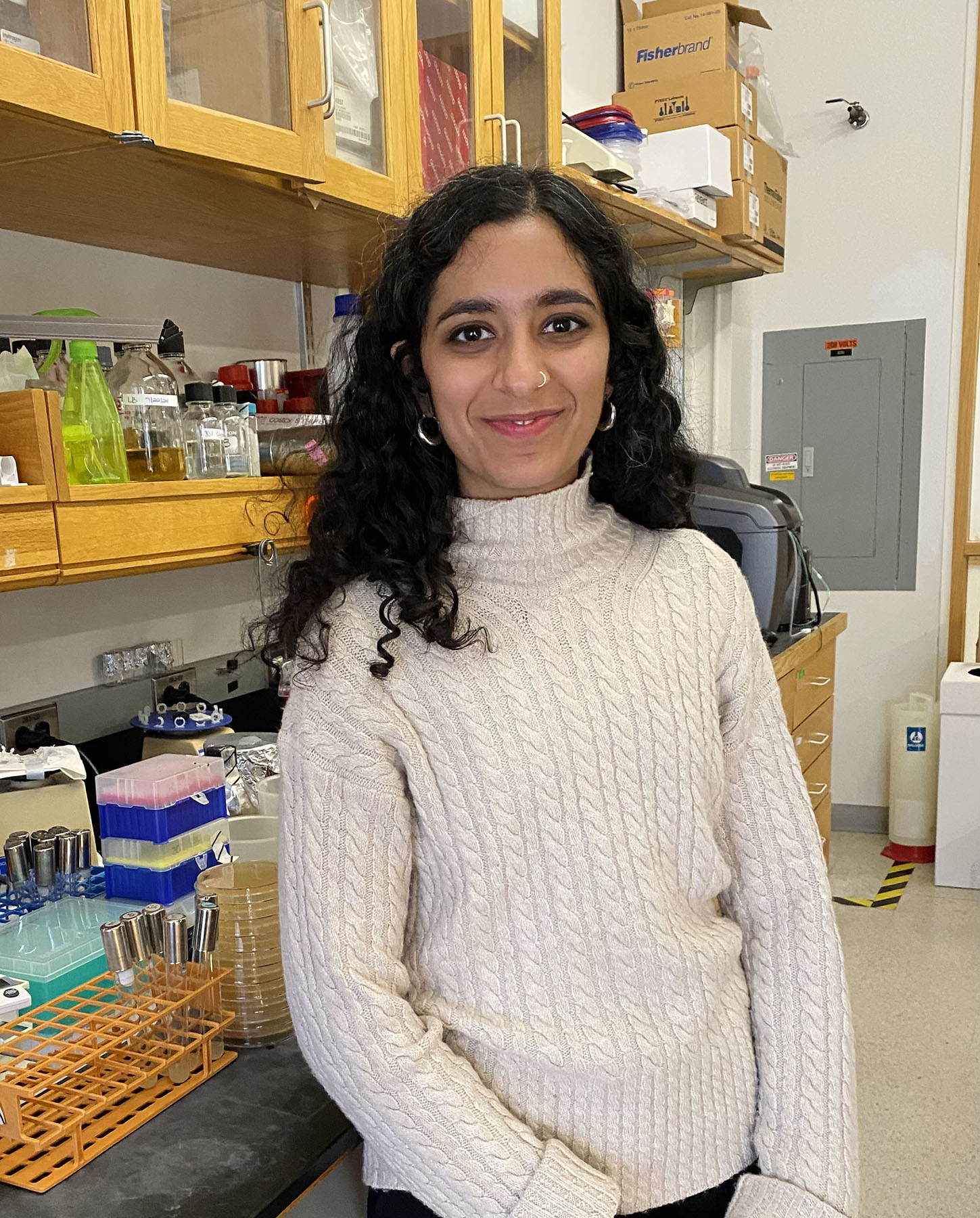
Dr. Gill [HHMI Fellow] is studying cell-cell communication via quorum sensing in developing biofilms. Biofilms are communities of bacteria that take on a three-dimensional structure and often develop striking visual features like wrinkles. Resident bacteria exploit this complexity to resist antimicrobial treatments and cause disease, particularly in healthcare settings, where biofilms pose serious threats to immunocompromised chemotherapy patients. Quorum sensing is the process by which bacteria orchestrate collective behaviors, including the assembly and dissolution of biofilm communities. Using quantitative microscopy at single-cell resolution, Dr. Gill is investigating how these signals between bacteria result in the construction of spectacular 3D biofilms at the air-liquid interface. A deeper understanding of how bacteria thrive in dynamic environments will contribute to new strategies to combat infections, which will positively impact treatments for all cancer types. Dr. Gill received her PhD from Harvard University, Cambridge and her BS from Drexel University, Philadelphia.
Dhiraj Indana, PhD
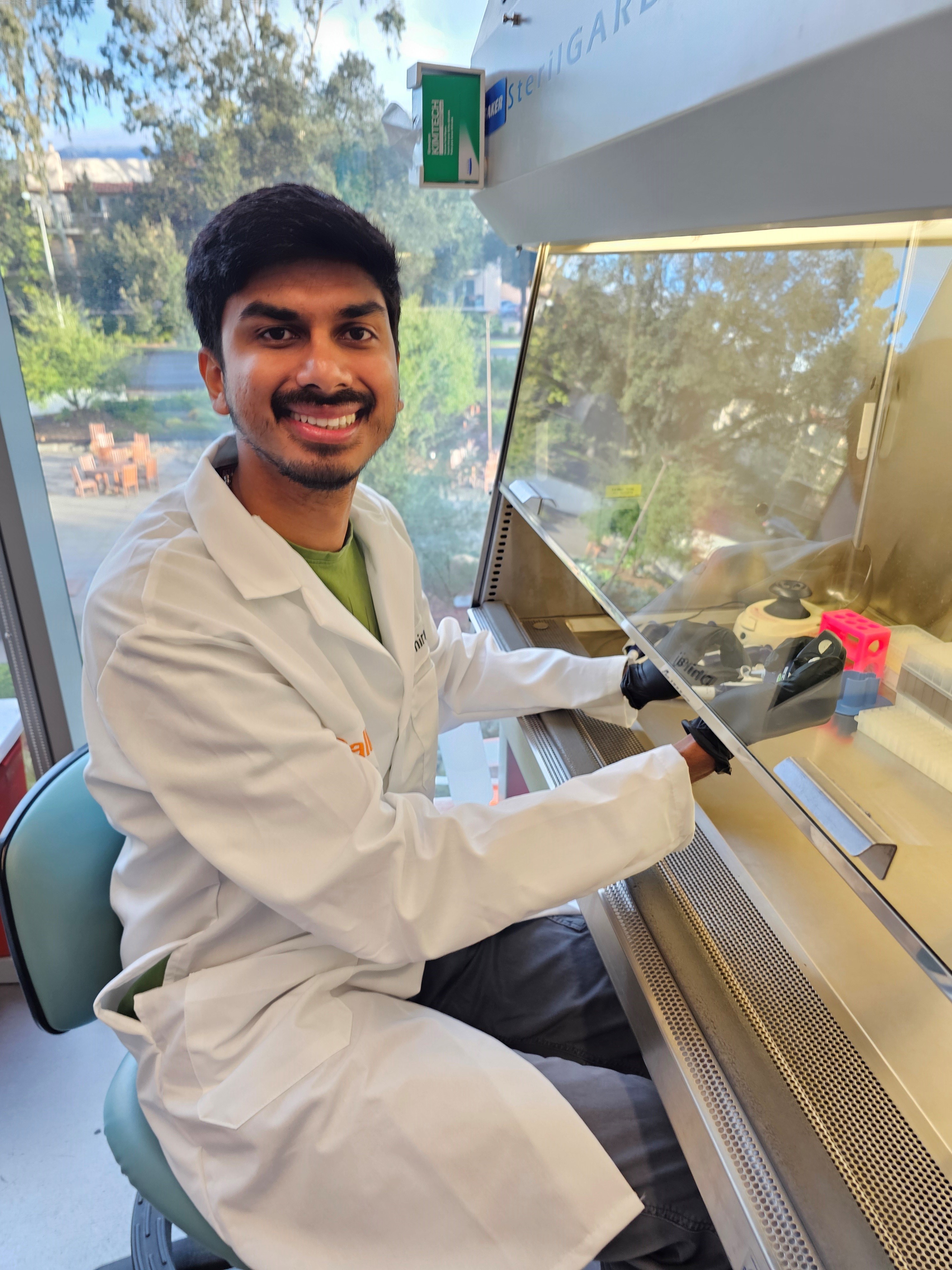
Successful immune responses against cancer require immune cells of various types to control each other’s proliferation, differentiation, and death. These interactions collectively constitute a set of intercellular signaling circuits. A fundamental challenge in cancer research is to understand the relationship between the architecture and functions of these circuits. Dr. Indana will quantitatively model and synthetically construct intercellular circuits with immune-like functions to understand how circuit architectures empower immune behaviors such as threat detection and cancer elimination while maintaining the ability to return to a non-inflammatory state. This work promises to uncover the “design principles” underlying various immune functions, providing a foundation for engineering novel immunotherapies. Dr. Dhiraj received his PhD and MS from Stanford University, Stanford and his BTech from Indian Institute of Technology, Roorkee.
Brendan Floyd, PhD
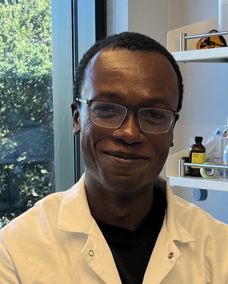
Proteins found on the surface of cells are key agents in cancer progression, as they play a role in cell signaling and metastasis. Targeted protein degradation has emerged as a therapeutic strategy to modulate what are considered “undruggable” proteins. Specifically, lysosomal-targeting protein degradation (LTPD), which uses the cancer cell’s own degradation machinery to break down proteins, has demonstrated therapeutic potential. However, the proteins targeted for LTPD have been limited to a few well-studied membrane and extracellular proteins, leaving much still unknown about the breadth of proteins that can be targeted for degradation and the features of a target protein that determine LTPD efficacy. Dr. Floyd [HHMI Fellow] aims to systematically characterize the features of cell surface proteins that drive the efficacy of LTPD with the goal of identifying new targets for blood cancer treatment. Dr. Floyd received his PhD from University of Texas at Austin, Austin and his BS from California Polytechnic State University, San Luis Obispo.
Ariën Schiepers, PhD
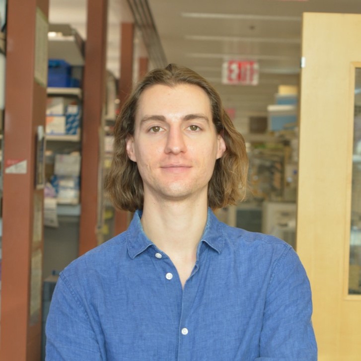
T lymphocytes, an important component of the immune system, recognize infected or cancerous cells with great specificity, ensuring targeted elimination. These potent cells are kept in check by regulatory T cells, the guardians of the immune system. While essential for curtailing excessive inflammation and preventing autoimmunity, their immunosuppressive properties can promote the development and progression of cancer. Regulatory T cells are distinguished by the presence of a protein called Foxp3, which plays a critical role in their differentiation, function and fitness. Foxp3 deficiency results in fatal autoimmune inflammatory disease, underscoring its importance for maintaining organismal health. Despite its significance, however, the reliance of regulatory T cells on Foxp3 in disease contexts like infection and cancer remains incompletely understood. Dr. Schiepers [HHMI Fellow] will study the fate and function of regulatory T cells in these settings using mouse genetics approaches and disease models of melanoma and colorectal cancer. Dr. Schiepers received his PhD from The Rockefeller University, New York and his MS and BS from Utrecht University, Utrecht.
Antonio J. LaPorte, PhD
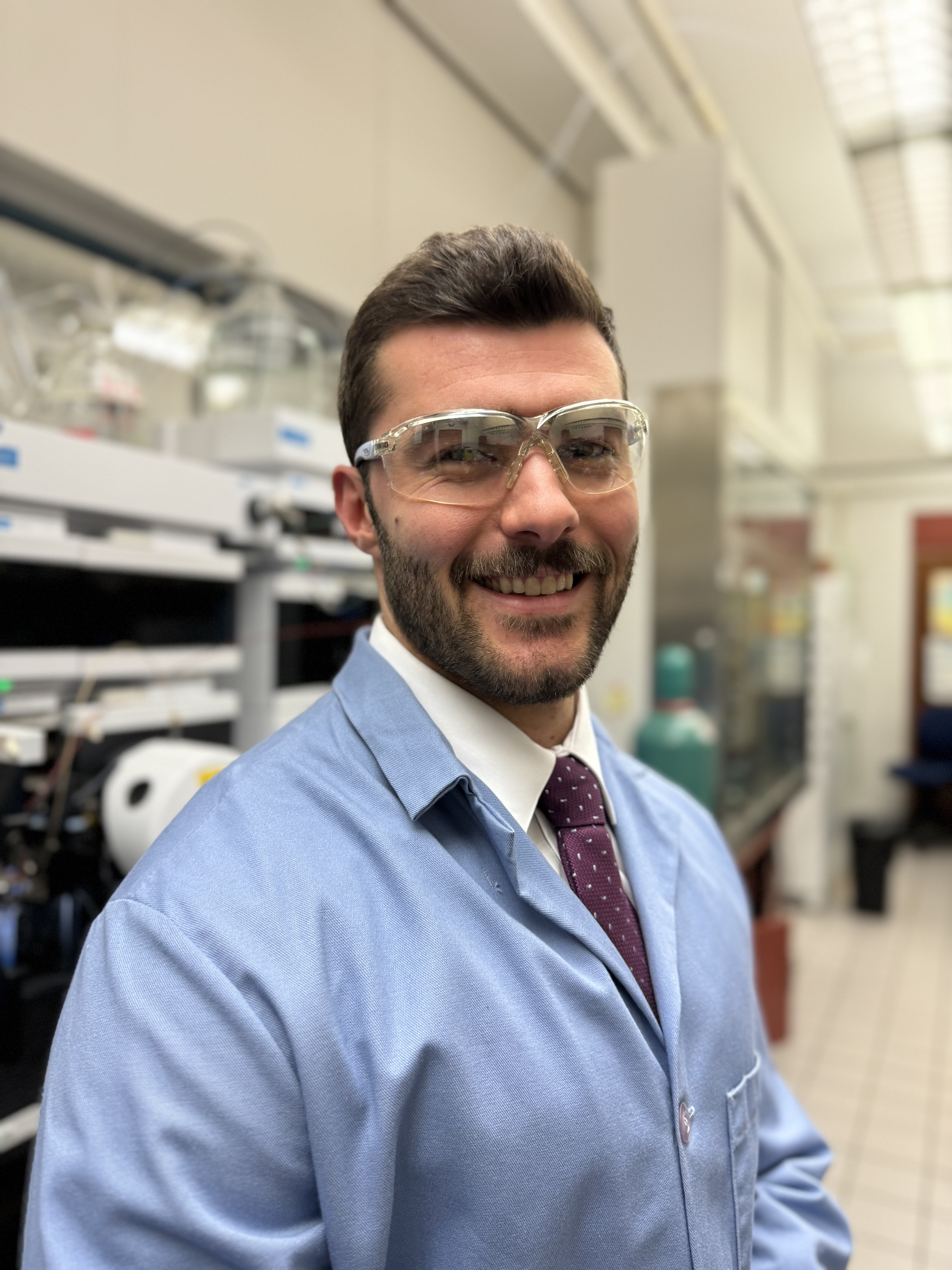
Sugar molecules on the surface of cells can be arranged in many different patterns, and it is the exact structure of a sugar that gives it its unique function. In sugars on the surface of cancer cells, the location of sulfate (SO4-) groups changes as the tumor grows and spreads. The study of sulfated sugars has been stymied, however, by the difficulty of synthesizing them in the laboratory. Dr. LaPorte’s research aims to remove this roadblock by developing a chemical reaction that easily constructs biologically important sugars with a sulfate group bound to them. These sulfated sugars will then be used to test how specific sulfation patterns affect their interaction with other biological proteins. The results of these experiments will lay the groundwork for new cancer treatments that use complicated sugar structures to selectively target cancer cells without harming healthy cells. Dr. LaPorte received his PhD from University of Illinois, Urbana-Champaign, Champaign and his BS from North Central College, Naperville.
Antonio Cuevas-Navarro, PhD
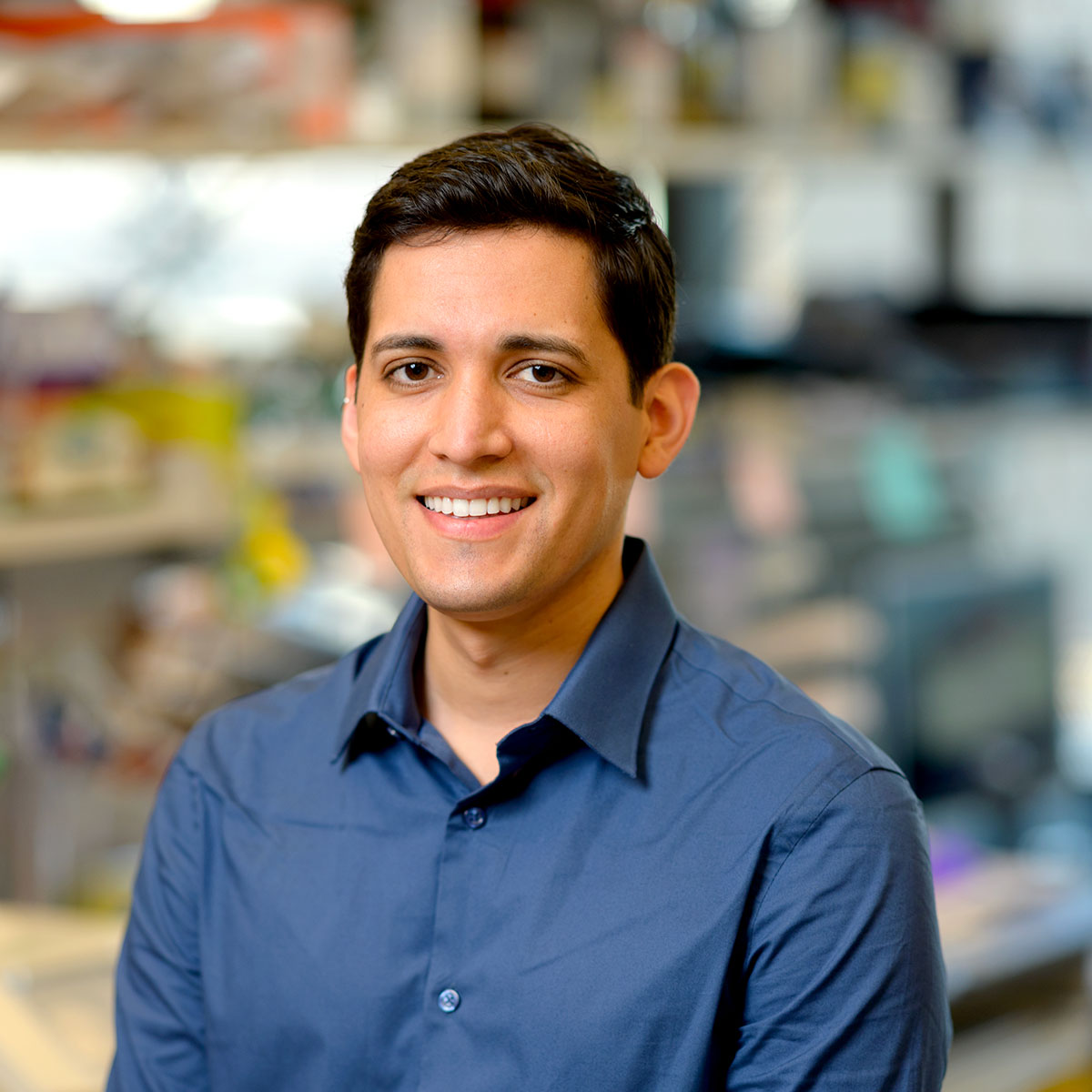
Dr. Cuevas-Navarro’s [Berger Foundation Fellow] research project focuses on targeting mutations in the RAS genes (HRAS, NRAS, and KRAS), present in about 30% of cancer patients and notorious for driving aggressive tumor growth. Dr. Cuevas-Navarro aims to mitigate these mutations’ effects by using pharmacological agents to enhance a biochemical process that regulates RAS proteins. His project will investigate the mechanism of action of these compounds and assess their effectiveness in patient-derived cancer models. This research has the potential to expand treatment options across various cancer types, including those where current treatments are limited. Dr. Cuevas-Navarro received his PhD from University of California, San Francisco and his BS from University of California, Davis.
Anita Reddy, PhD
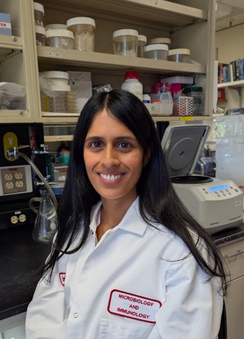
The Western diet, characterized by low dietary fiber and high saturated fat content, is strongly correlated with incidence of CRC. This diet rapidly alters the composition and function of the gut microbiome, inducing microbial imbalance and inflammation, two major hallmarks of CRC. These changes are thought to be caused by diet-induced oxygenation of the gut environment. Most beneficial gut bacteria cannot survive this oxygenated environment, but harmful microbes can thrive in this setting and further exacerbate inflammatory responses. The response of bacteria to oxygen is varied and poorly understood. Dr. Reddy [Connie and Bob Lurie Fellow] aims to elucidate the mechanism underlying oxygen tolerance in bacteria and find interventions to restore normal oxygen levels. The findings from this project will delineate the contributions of exercise and diet to establishing the oxidative environment of the gut and illuminate the mechanism allowing certain microbes to withstand redox stress. Dr. Reddy received her PhD from Harvard University, Cambridge and her BS from University of Connecticut, Mansfield.
Sagar Bhattacharya, PhD
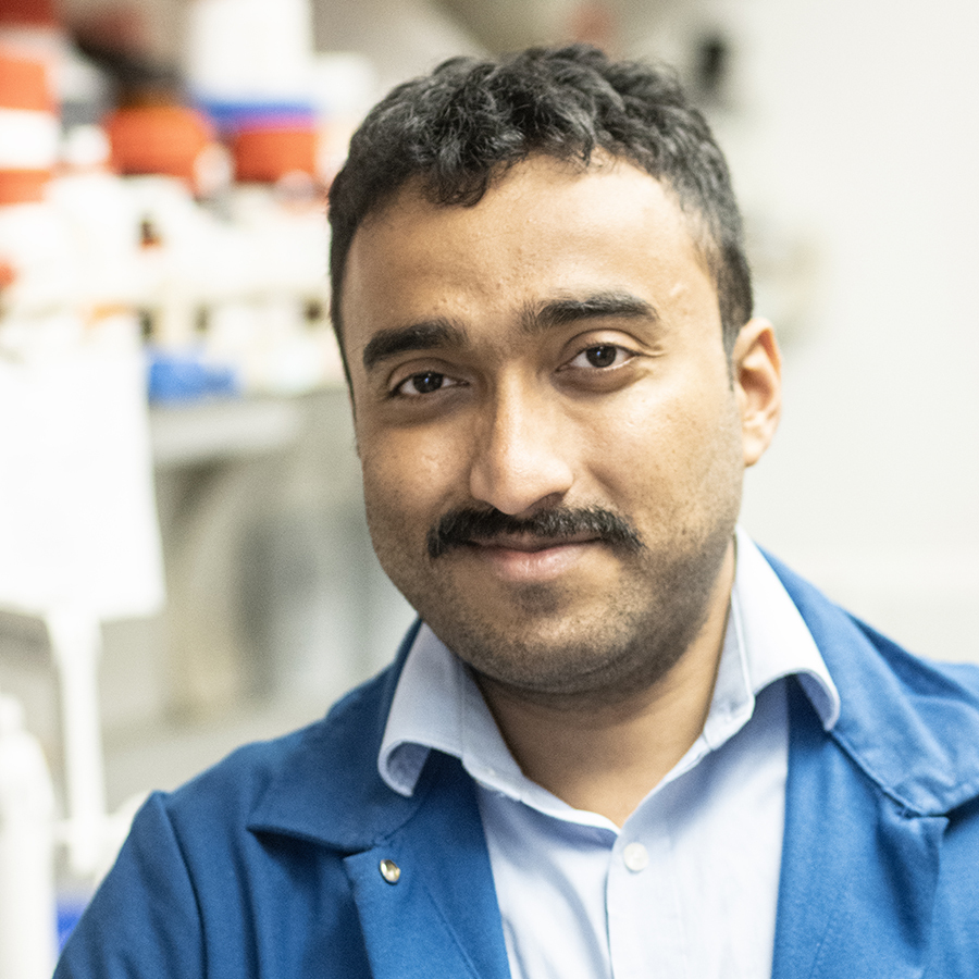
Peptide drugs, which mimic the function of natural peptides such as hormones or growth factors, have emerged as a promising strategy for the treatment of cancer. Despite their potential, however, very few have reached the clinic in the past decade, primarily due to their off-target toxicity. The design of a suitable system to deliver peptides in a site-specific manner would address a major challenge in the development of anticancer peptide drugs. De novo protein design, or building proteins “from scratch,” has allowed for the engineering of functional proteins for a broad range of applications, from catalysis to pharmaceuticals. Dr. Bhattacharya [Connie and Bob Lurie Fellow] aims to design proteins from scratch that can “mask” a peptide of interest for systemic delivery to the desired location. This project will initially target pediatric sarcomas, but eventually extend to other cancers like glioblastoma and breast cancer. Dr. Bhattacharya received his PhD from Syracuse University, Syracuse and his MS and BS from University of Calcutta, Kolkata.
Shaohua Zhang, PhD
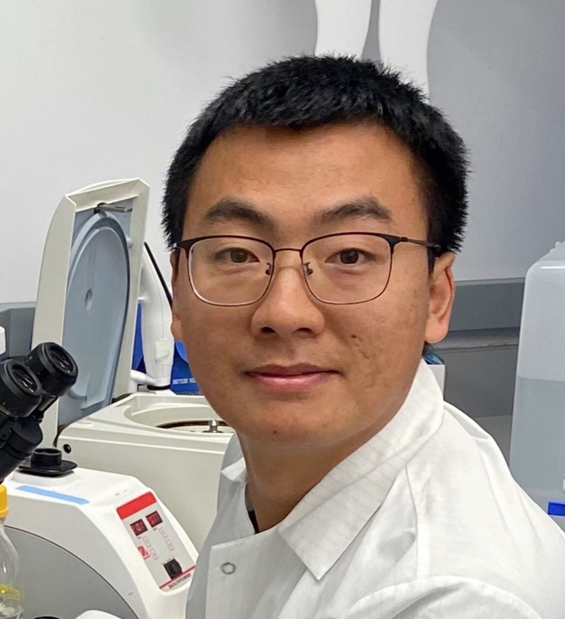
Dr. Zhang [Timmerman Traverse Fellow] aims to engineer T cells with synthetic cell adhesion molecules (synCAMs) to augment current approaches for immunotherapy. This project represents a fundamentally new strategy for CAR T cell engineering that could overcome tumor escape from immunotherapy across multiple forms of cancer. Understanding how synCAMs contribute to CAR T cell efficacy will provide insights beyond cytotoxic CAR T cell therapy; this work could lead to the application of synCAMs in other engineered immune cell therapies under investigation, such as CAR macrophages, CAR natural killer cells, and CAR T regulatory cells. Overall, this approach could lead to CAR T cells that are much more robust to tumor evasion and target antigen expression, and thus much more effective therapeutically. Dr. Zhang received his PhD from University of Chinese Academy of Sciences, Shanghai and his BS from Wuhan University, Wuhan.
Lucia Ichino, PhD
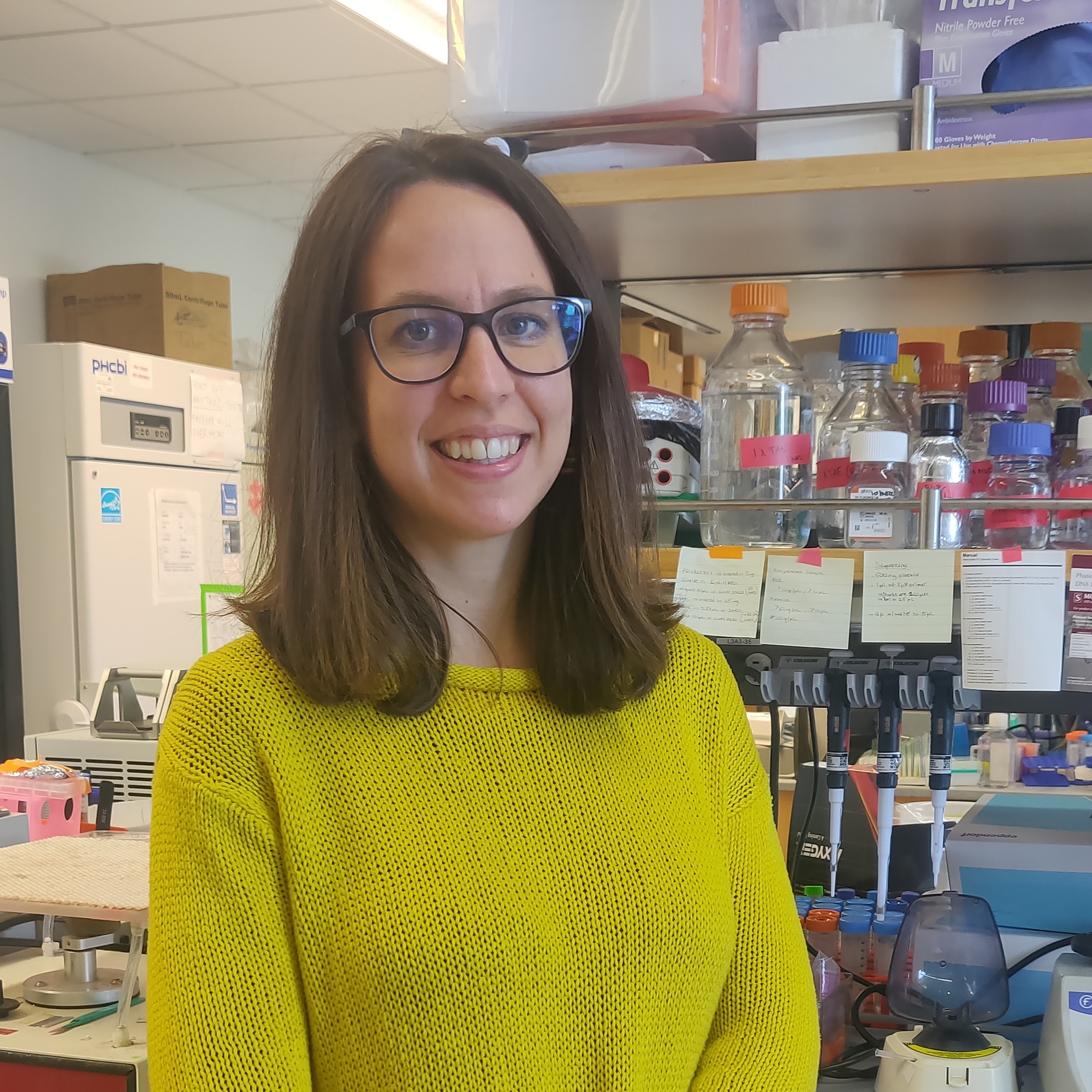
Epithelial to Mesenchymal Transition (EMT) is a crucial biological process that occurs during early development. It allows epithelial cells, which line the inner and outer surfaces of the body, to undergo a profound transformation in cellular identity and migrate and populate the embryo. Unfortunately, numerous cancer types exploit this mechanism, allowing cancer cells to detach from the tissue of origin and disseminate throughout the body, significantly worsening patients’ prognoses. Dr. Ichino [HHMI Fellow] is studying the process of developmental EMT with the goal of discovering novel ways to interfere with it in the context of cancer progression. Dr. Ichino’s research takes advantage of a lab-grown system that mimics the EMT and migration of neural cells. Using this system, she plans to study how EMT-promoting transcription factors orchestrate this global change in cellular identity, and how genetic variations can influence this process. Dr. Ichino received her PhD from University of California, Los Angeles and her MS and BS from San Raffaele University, Milan.
Brooke D. Huisman, PhD
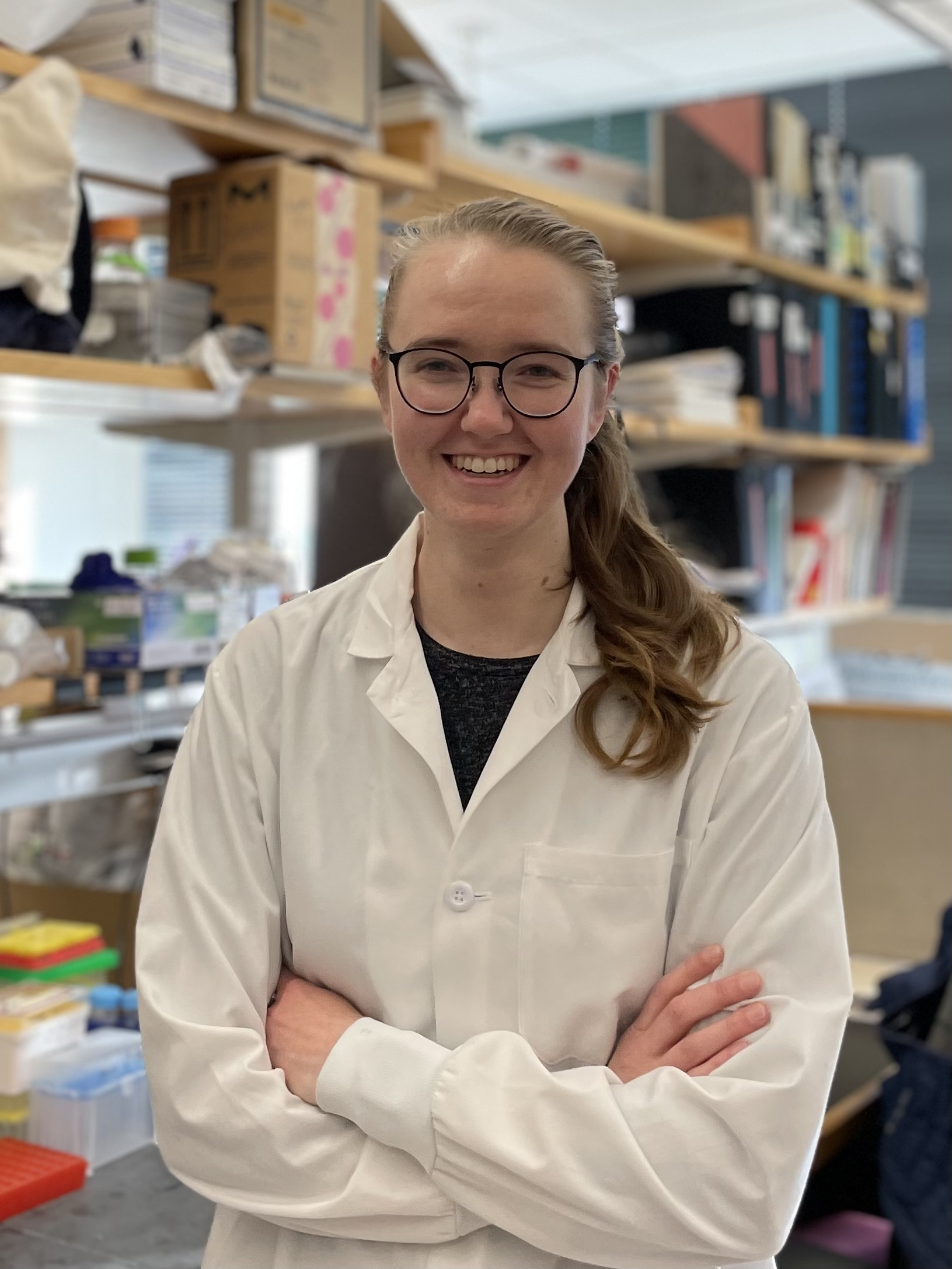
To prevent autoimmune attacks, T cells are screened in the thymus to ensure they do not react to self-derived antigens. Dr. Huisman [National Mah Jongg League Fellow] studies the thymus and, specifically, a population of cells called “thymic mimetic cells” that mimic other tissues, such as muscle or gut, and assist T cells in developing tolerance to diverse cell types. Dr. Huisman’s research focuses on understanding how thymic mimetic cells develop. This work may lead to improved understanding of thymus-mediated tolerance to tumors, novel therapeutic opportunities for manipulating mimetic cells to induce anti-tumor responses, and increased understanding of thymic tumors. Dr. Huisman received her PhD from Massachusetts Institute of Technology, Cambridge and her BS from University of Michigan, Ann Arbor.
Cheng Yang, PhD
Protein oxidation occurs when an amino acid gains an oxygen atom in a post-translational modification. Oxidation of the amino acid methionine plays an important role in cellular regulation, and mutations at methionine sites are known to have pathogenic effects in cancer. However, direct assessment of methionine’s oxidation product, methionine sulfoxide, remains underexplored. Dr. Yang aims to develop methionine sulfoxide labeling approaches using light or electricity. With the help of these chemical tools, he will profile and identify methionine sulfoxide sites in pancreatic tumors and study their role in metastasis. Dr. Yang received his PhD from the University of Michigan, Ann Arbor and his BS from Nankai University, Tianjin.
Jinchun Wu, PhD
Genome rearrangements have been widely observed in human cancers. Recent whole-genome sequencing data has identified chromothripsis, an event that introduces massive genome rearrangements in only one or a few chromosomes through catastrophic shattering and random reattachment, as one of the most frequent genome rearrangements. Chromothripsis has been associated with poor clinical outcomes in multiple cancers, but the shattering mechanisms that induce chromosome fragmentation remain uncharacterized. Dr. Wu [Marion Abbe Fellow] aims to determine the role of cytoplasmic nucleases (enzymes that cleave DNA) in chromosome shattering and genome rearrangement, which will contribute to our understanding of chromothripsis in all cancers. She will extend this project to a mouse model of glioma to determine the effects of candidate nucleases on cancer progression. Dr. Wu received her PhD and BS from Peking University, Beijing.
Simon Sretenovic, PhD
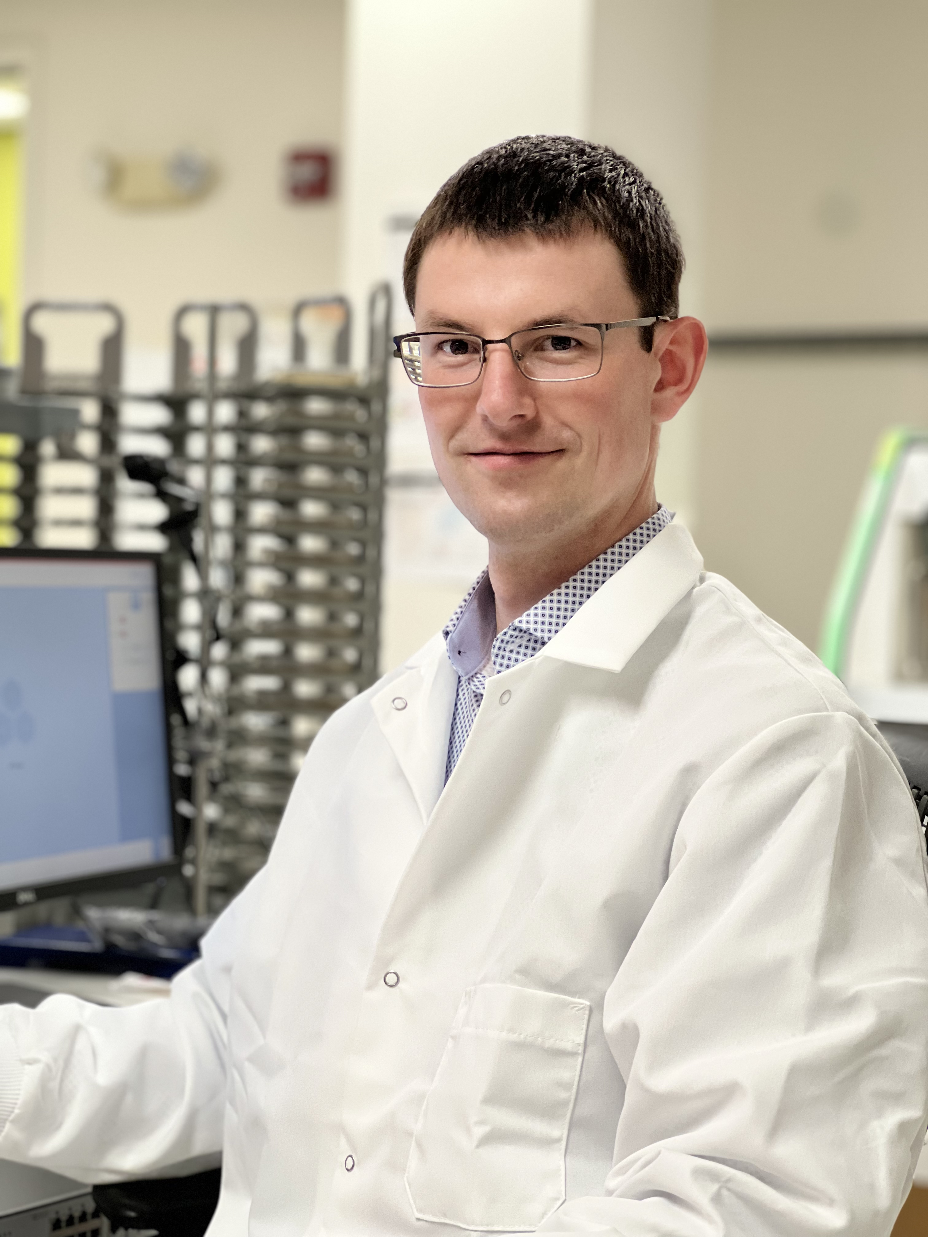
More than one third of all people will receive a cancer diagnosis at some point in their lifetime. Dr. Sretenovic [Connie and Bob Lurie Fellow] is using both yeast and human cell lines to model various properties of cancerous cells as complex genetic traits. Combining novel CRISPR genome editing approaches with next-generation sequencing technology, he aims to dissect the intricate relationships between genetic variants, chemical and physical environmental factors, and phenotypic outcomes (i.e., observable characteristics). The goal of his project is to understand the genetic basis for a panel of cancer-related traits to inform the development of anti-cancer treatments. Dr. Sretenovic received his PhD from the University of Maryland, College Park, and his MS and BS from University of Ljubljana, Ljubljana.
Wenzhi Song, PhD
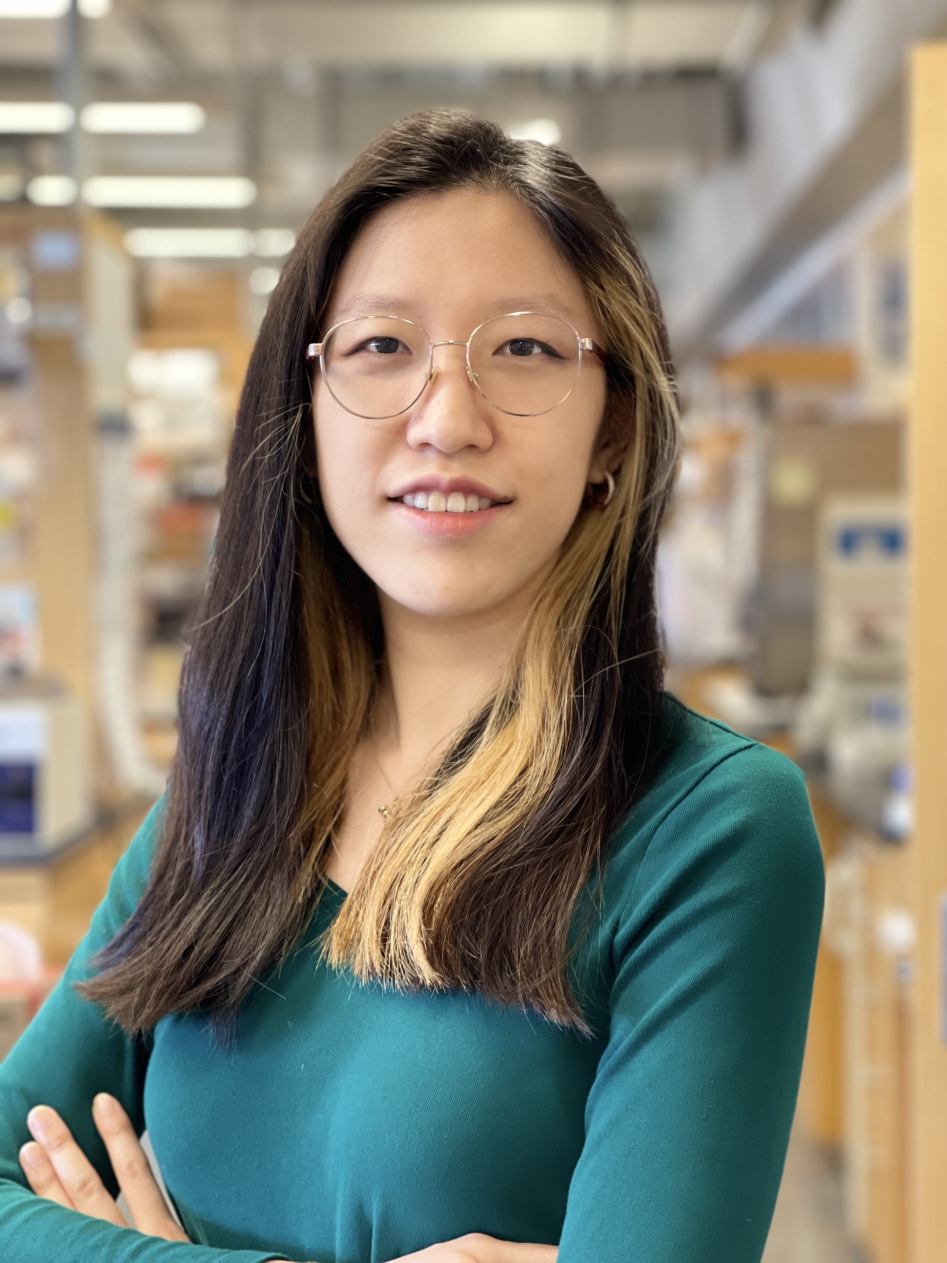
The interaction between cancer cells and their non-malignant neighbors in the tumor microenvironment is critical for cancer progression. While certain types of cellular crosstalk within the tissue safeguard against malignancy, cancer cells are often able to exploit nearby cells to fuel tumor growth. Dr. Song [HHMI Fellow] is interested in understanding how the complex cellular communication network in the skin, namely its sensory and immunological components, contributes to the development of cutaneous squamous cell carcinoma, one of the most common skin cancers. Identifying novel neuronal and immunological interactions within the tumor microenvironment has the potential to uncover pathways regulating cancer progression and anti-tumor immunity. Dr. Song received her PhD from Yale University, New Haven and her AB from Bryn Mawr College, Bryn Mawr.
Yoshiki Sakai, PhD
Normally, epithelial tissues, which cover all external body surfaces and line internal cavities, expel unwanted cells to maintain health in a process known as cell extrusion. However, some cancer cells, particularly those with the common RasV12 mutation, manage to avoid extrusion. Using Drosophila (fruit flies) as a model, Dr. Sakai [Rhee Family Fellow] will explore how RasV12-mutant cells manipulate neighboring cells to avoid extrusion. Understanding this process could lead to new ways to prevent cancer cells from escaping the epithelial defense, offering potential new treatments. Dr. Sakai received his PhD and BS from Nagoya University, Nagoya.
Ian J. Roney, PhD
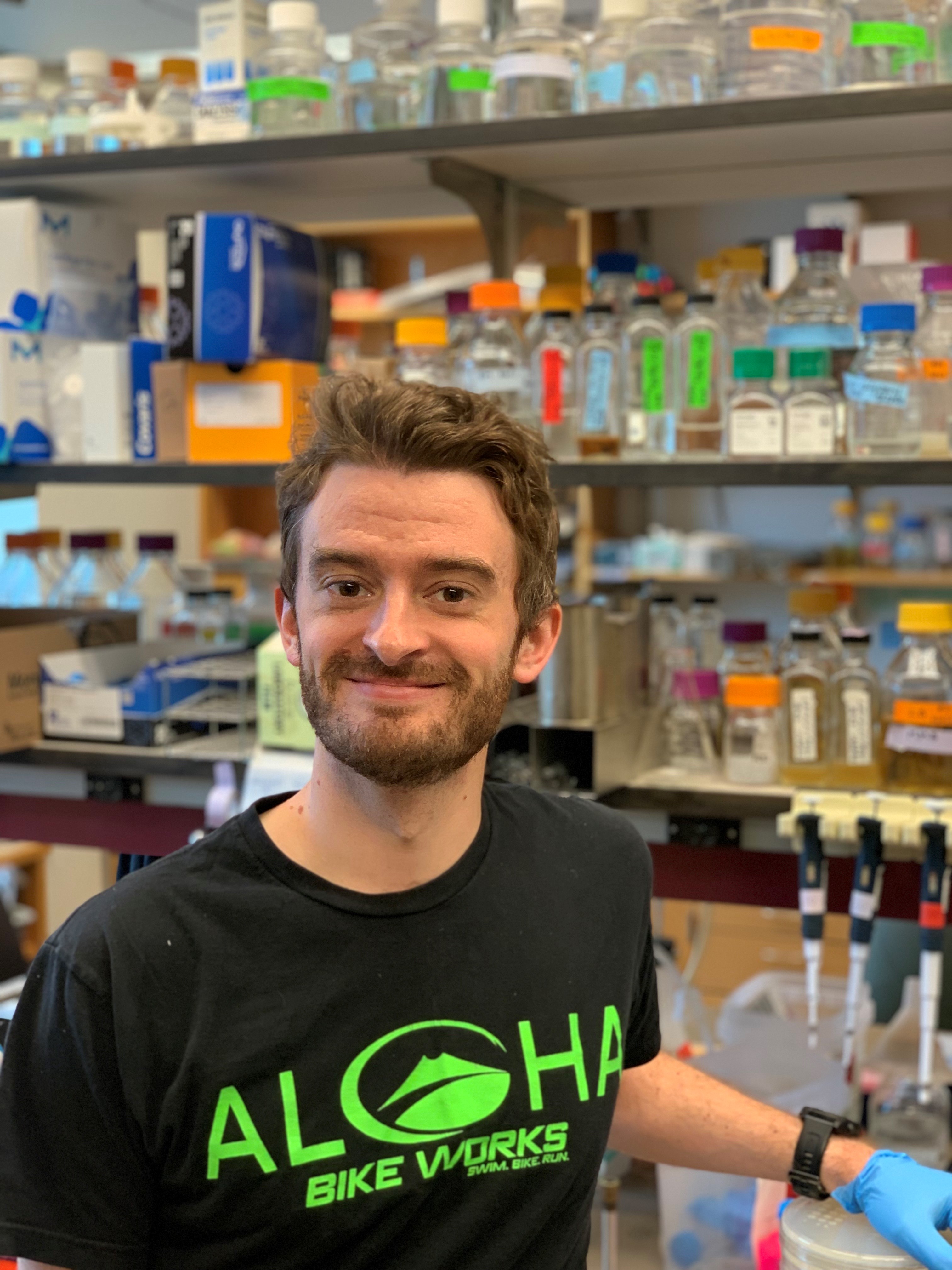
Bacteria have diverse immune systems to defend themselves against viral invaders, many of which use molecular mechanisms also seen in mammalian immune systems. Dr. Roney [HHMI Fellow] studies how bacterial immune systems detect virally compromised cells, and how viruses undermine immune systems to prevent the elimination of virally compromised cells from the population. The goal of his research is to uncover novel mechanisms and principles of immune systems that are found across domains of life. The discoveries resulting from this work will broaden our understanding of how immune systems detect and eliminate compromised cells, like cancer cells, and could help guide development of new immunotherapies. Dr. Roney received his PhD from Harvard University, Cambridge and his MS and BS from University of Ottawa, Ottawa.
David S. Roberts, PhD
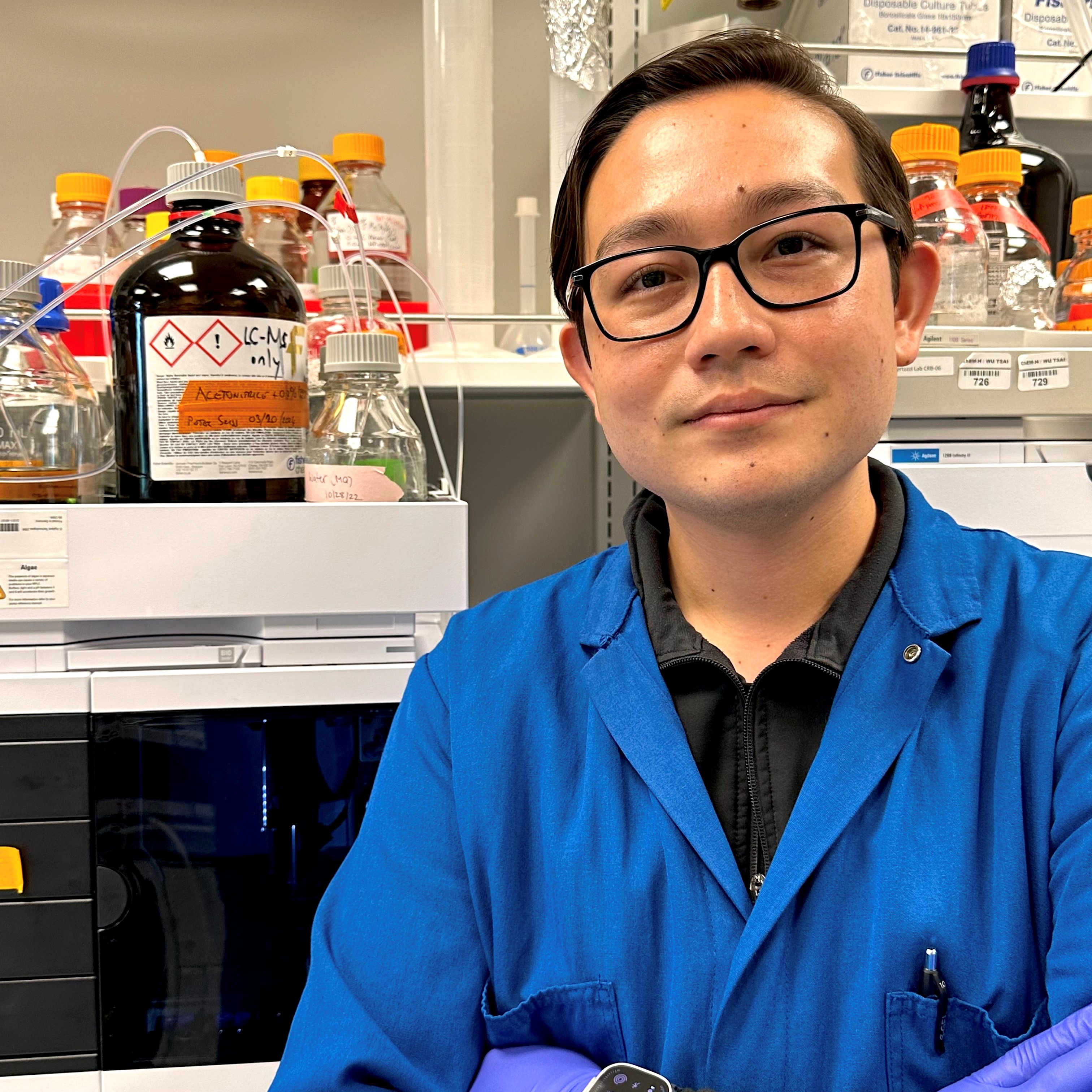
Cancer immunotherapies have shown remarkable benefits, but many tumors remain unresponsive to existing treatments. The mechanisms cancer cells use to evade immune responses during treatment remain largely unknown. Altered cell surface glycosylation, the process of attaching sugars to cell surface biomolecules, is a hallmark of many human cancers. The interaction between cell surface glycoproteins on immune cells with cancer cells represents a major axis of immune evasion and plays a vital role in how cancer cells suppress immune responses during cancer treatment. Dr. Roberts’ [Connie and Bob Lurie Fellow] research aims to molecularly define cell surface glycosylation and understand the role of glycosylation in driving cancer immunosuppression. This knowledge will be leveraged to illuminate the underlying mechanisms of tumor immune evasion and enable next-generation classes of cancer immunotherapies. Dr. Roberts received his PhD from University of Wisconsin–Madison, Madison and his BS from University of California, San Diego.
Nalin Ratnayeke, PhD
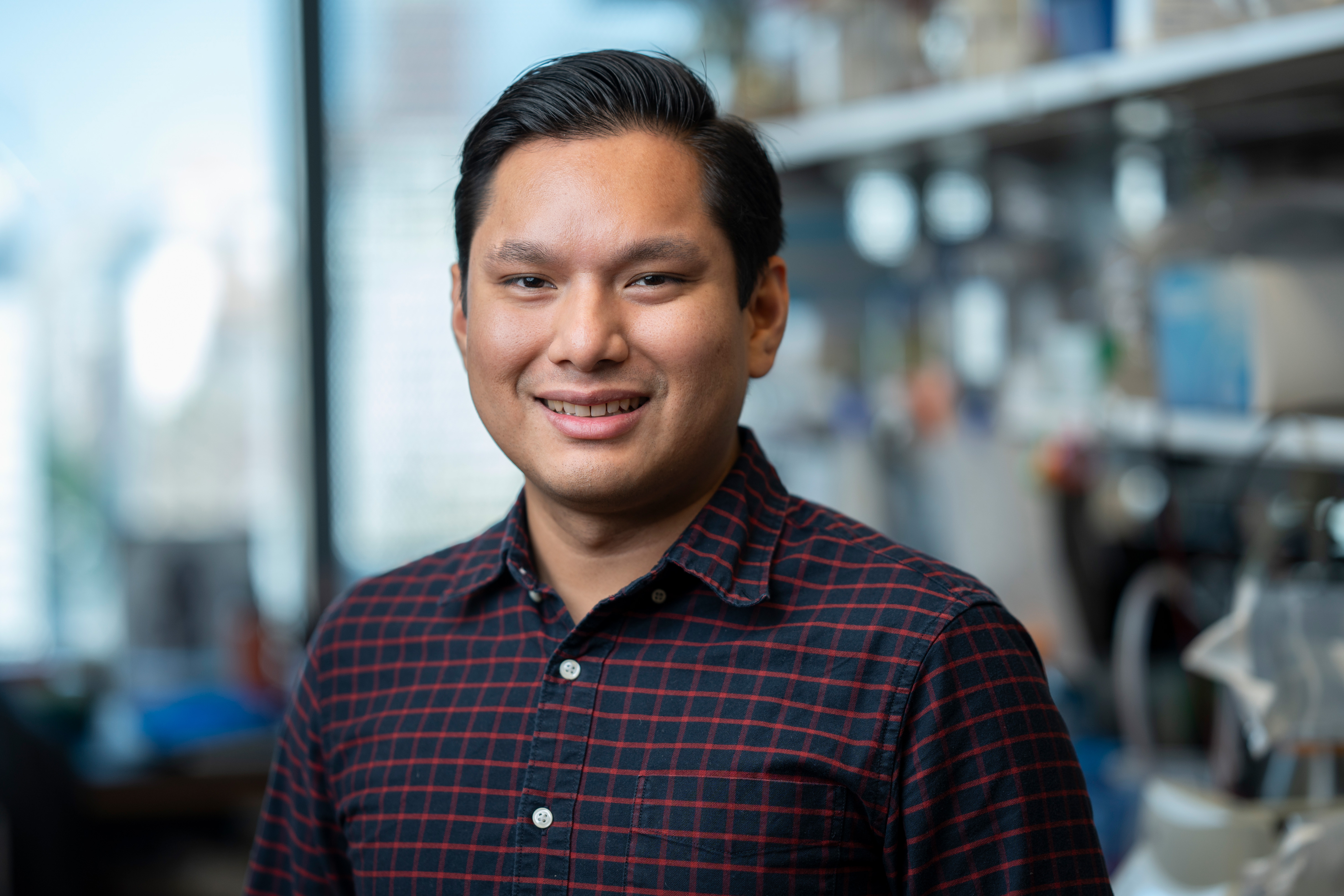
Pancreatic cancer is a leading cause of cancer-related deaths. The development of drugs targeting mutant KRAS, the oncogenic driver of most pancreatic cancers, has led to much optimism for improved treatments. However, tumor recurrence driven by heterogeneous cancer cell responses to these drugs remains a major challenge. Some cancer cells die, while surviving cells can halt their proliferation or continue to proliferate in the presence of drug, all of which can occur within the same tumor and dictate the overall response to treatment. Dr. Ratnayeke [HHMI Fellow] is studying the mechanisms that underlie these heterogeneous responses using mouse models of pancreatic cancer and single-cell genomics to map cellular states to their drug responses. Understanding these mechanisms will inform combination and precision therapies with mutant KRAS-targeting drugs to tune tumor responses in beneficial directions. Dr. Ratnayeke received his PhD from Stanford University, Stanford and his BS from the University of Texas at Austin, Austin.
Sarah L. Price, PhD
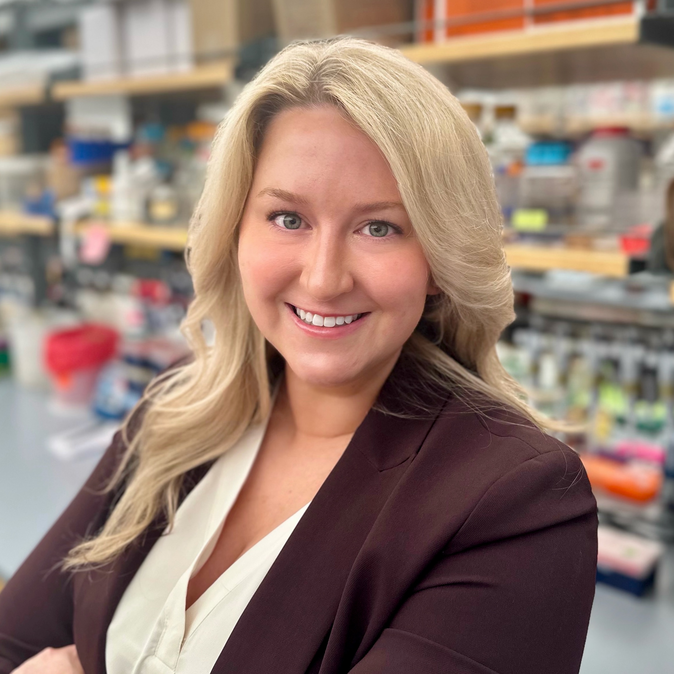
Emerging evidence implicates the pathogenic bacterium C. difficile as an initiator of colorectal cancer. C. difficile exposure can lead to chronic recurrent disease that is difficult to clear with antibiotics. The generation of spores is a well-studied mechanism used by C. difficile to persist; however, other mechanisms of recurrent infection remain poorly understood. Dr. Price [Merck Fellow] hypothesizes that biofilms may function as reservoirs of C. difficile and aims to elucidate their role in disease relapse. She will employ innovative imaging strategies to visualize the composition and development of C. difficile biofilms in the gastrointestinal tract, with the goal of generating insight that will improve treatments for C. difficile infections and identify strategies to prevent colorectal cancer. Dr. Price received her PhD from University of Louisville, Louisville and her BS from University of Tennessee, Knoxville.
Sangwoo Park, PhD
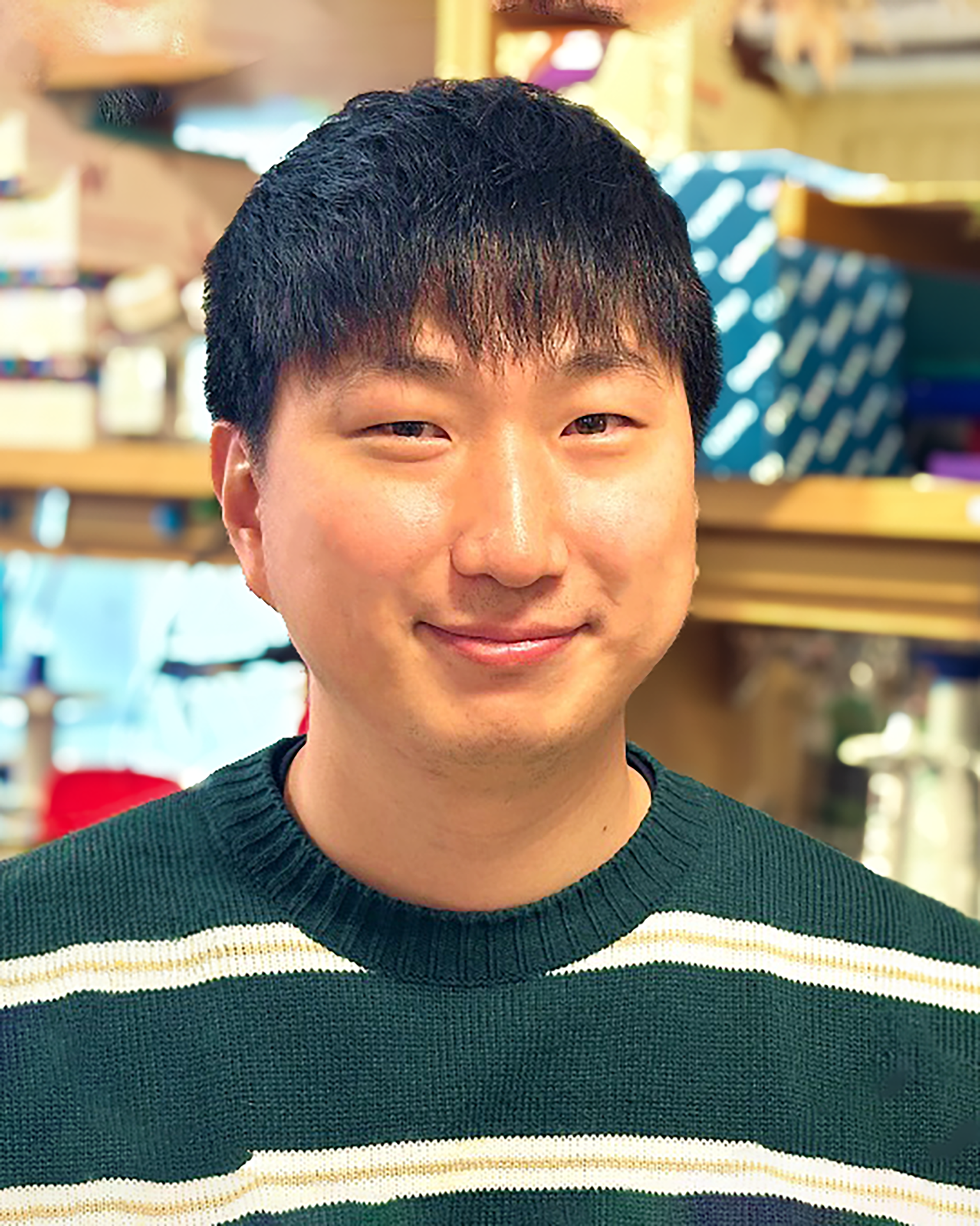
One way cancer cells evade immune attack is by constructing a thin material barrier called the glycocalyx on their surface to evade detection and destruction by surveilling immune cells. Tiny changes in the glycocalyx thickness, as small as 10 nanometers, can affect the anti-tumor activity of immune cells, including CAR T cells. Dr. Park’s [Merck Fellow] goal is to develop strategies to endow CAR T cells with the ability to penetrate the glycocalyx barrier in solid tumors such as breast cancer and glioblastoma. These strategies will increase the effectiveness of CAR-T cell therapy against solid tumors by overcoming a significant mechanism of immune cell evasion. Dr. Park received his PhD from Cornell University, Ithaca and his BS from Korea Advanced Institute of Science and Technology, Daejeon.
Expery O. Omollo, PhD

Dr. Omollo [Robert A. Swanson Family Fellow] studies how bacteria have evolved to achieve precise gene expression using strategically placed transcription terminators. In cancer cells, specific mutations lead to uncontrolled transcription of certain genes, resulting in elevated gene expression that fuels cancer progression. Using bacteria as a model, Dr. Omollo aims to uncover how RNA polymerases in cancer cells evade termination signals to maintain high levels of gene expression, encouraging cancer spread. Dr. Omollo received his PhD from University of Wisconsin-Madison, Madison and his BS from Michigan State University, Lansing.
Chaiheon Lee, PhD
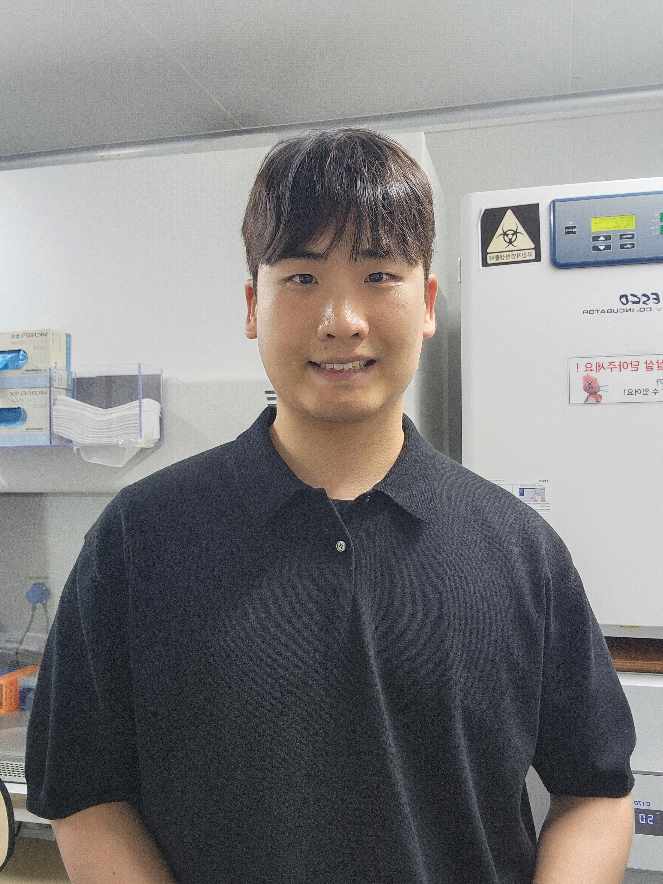
The immune system has the capability to destroy cancer cells harboring mutated genes. Cells display peptides derived from these mutated genes (i.e., portions of the mutant protein) on a molecule called the major histocompatibility complex I (MHC I), triggering cytotoxic T cells to eliminate the cancer cells. Unfortunately, this surveillance system is weak and often subverted by cancer cells. Dr. Lee [Suzanne and Bob Wright Fellow] aims to enhance the immunogenicity of the MHC I-displayed peptides using haptens, small molecules that elicit an immune response when attached to a larger carrier protein. By empowering the immune system, he envisions that these hapten-protein complexes will enable the repurposing of cancer drugs for which resistance has emerged. Dr. Lee received his PhD and BS from the Ulsan National Institute of Science and Technology, Ulsan.
Michael V. Gormally, MD, PhD
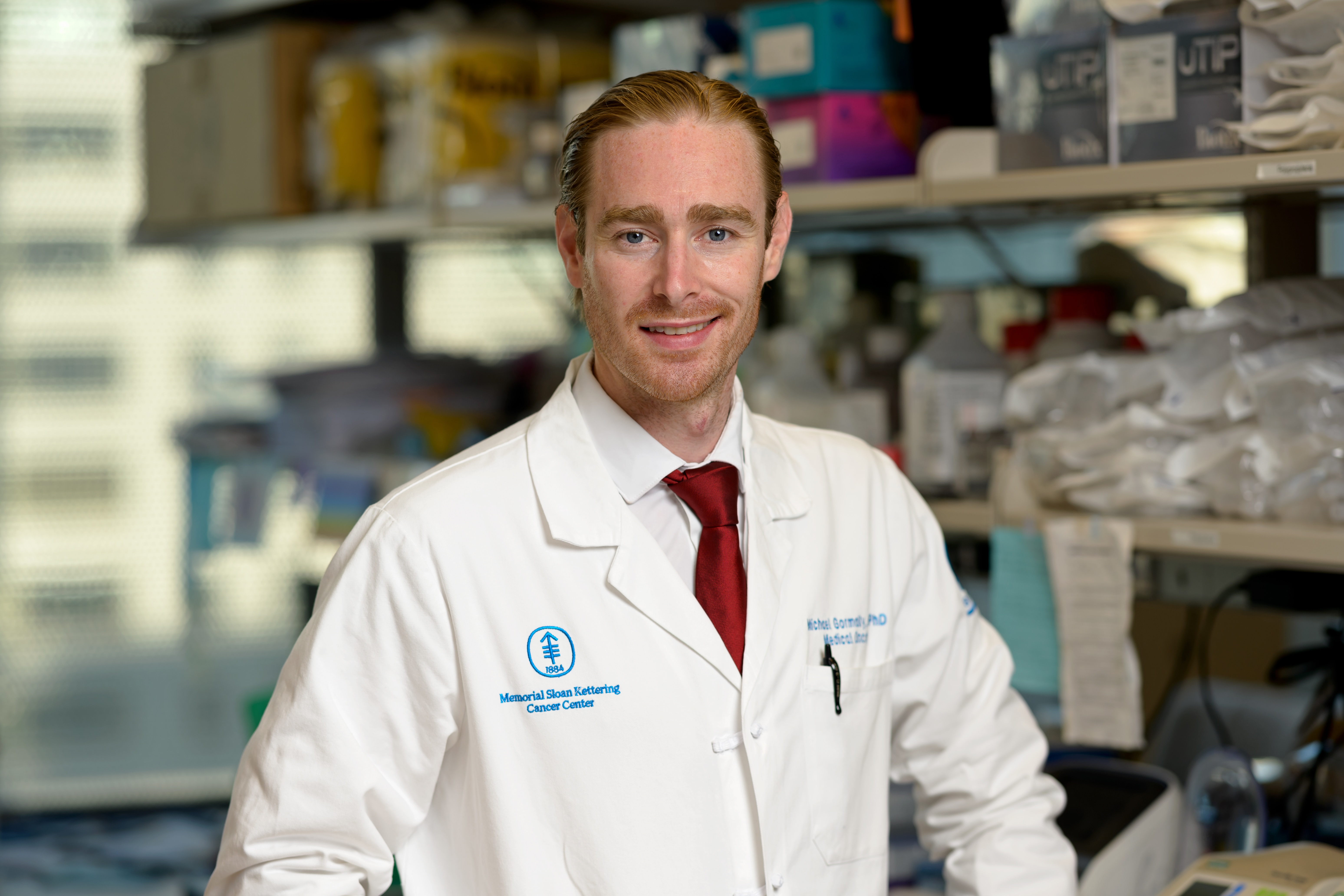
Adoptive cell therapy (ACT) is poised to expand the curative potential of immunotherapy. ACT works by administering T cells that have been genetically engineered to express tumor-specific T cell receptors (TCRs) so that they recognize a particular cancer antigen. Dr. Gormally’s [Dennis and Marsha Dammerman Fellow] work addresses two major challenges that currently limit the effectiveness of ACTs against solid tumors: identifying antigen targets that can be recognized by the immune system, and designing TCRs that target those antigens with exquisite specificity. Dr. Gormally and his colleagues have identified multiple immunogenic antigens derived from cancer-causing mutations and developed a powerful approach to retrieve potent, antigen-specific TCRs from large libraries of blood samples from cancer patients. The goal of these efforts is to identify safe and effective TCRs for clinical application. Dr. Gormally received his PhD from the University of Cambridge, Cambridge, his MD from Yale School of Medicine, New Haven, and his BA from Pomona College, Claremont.
R. Camille Brewer, PhD
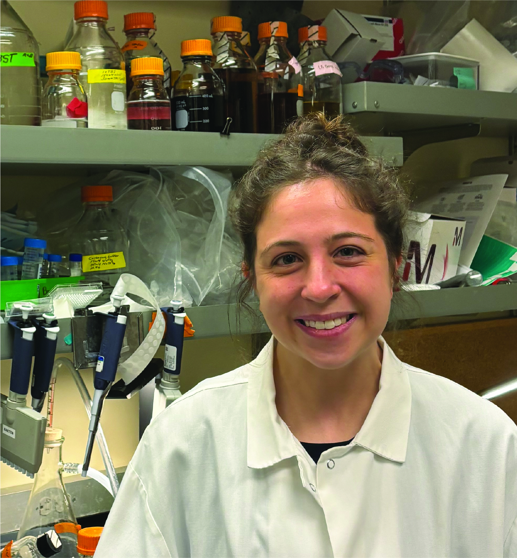
B cells, especially those that target cancer antigens, are crucial for fighting tumors; however, not everyone develops them. Our gut bacteria play a vital role in training B cells to recognize a wider range of threats. Dr. Brewer’s [HHMI Fellow] research explores how these gut bacteria influence the specificity of B cells, and thus our body’s ability to combat tumors. Dr. Brewer’s research aims to determine if the “training” of B cells by gut bacteria early in life influences their later responses to vaccines and cancer. This investigation may not only improve our understanding of how gut bacteria shape our immune system, but also pave the way for novel cancer treatments utilizing gut bacteria. Dr. Brewer received her PhD from Stanford University, Stanford and her BS from Massachusetts Institute of Technology, Cambridge.
Layla J. Barkal, MD, PhD
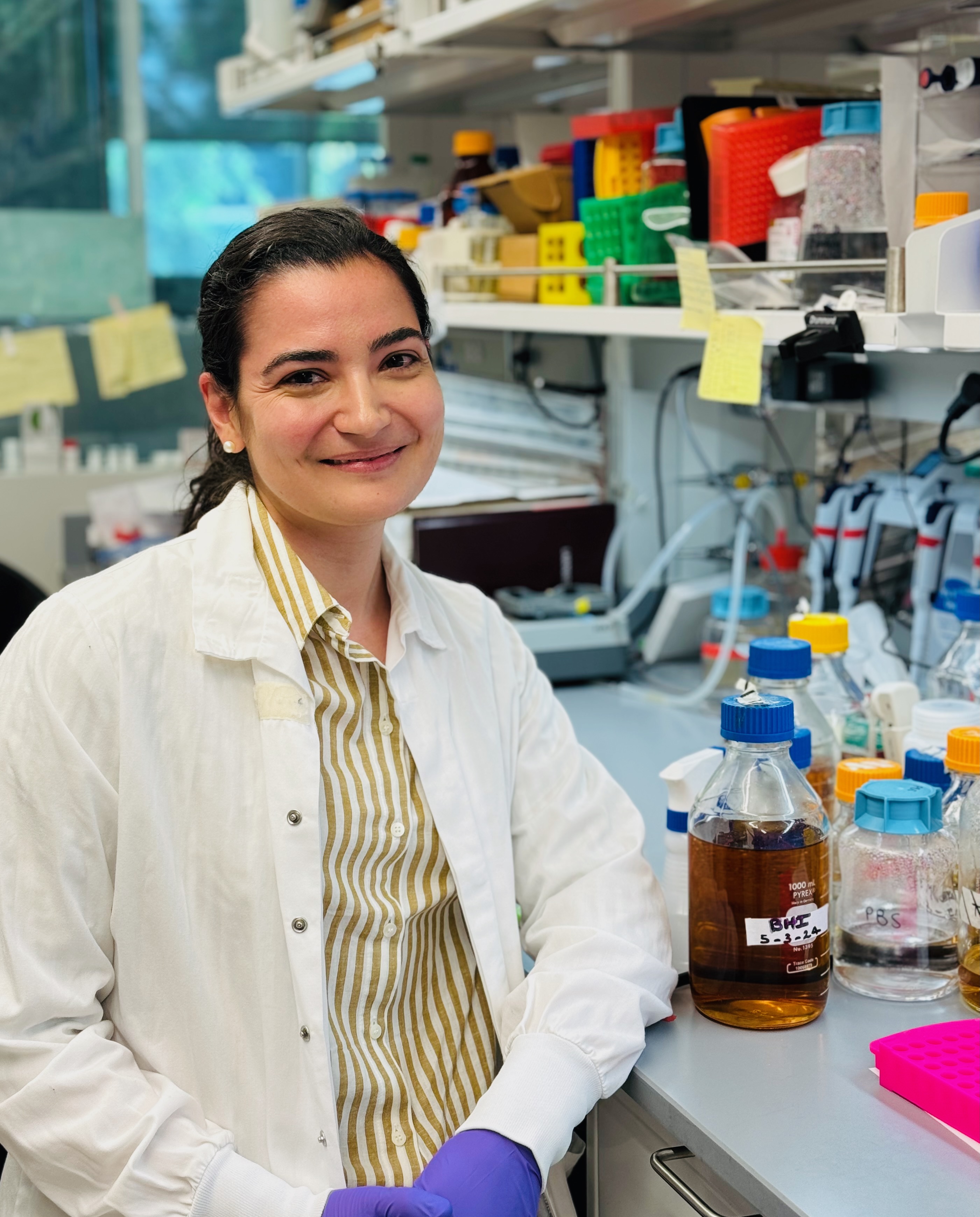
The bacterium Staphylococcus epidermidis (S. epi) is nearly universally present on human skin, and certain strains are capable of eliciting immune responses that can be redirected against tumor antigens. Dr. Barkal is investigating how to harness the immunomodulatory properties of S. epi to develop a new class of T cell immunotherapy that is potent and tumor antigen-specific, avoiding the systemic side effects associated with current immunotherapies. Specifically, she is using a melanoma model to explore how to modulate T cell production with S. epi and how to use other skin bacteria for synergistic anti-tumor effects. This work will form the foundation for human trials of topical bacteria-based cancer immunotherapy. Dr. Barkal received her MD, PhD from University of Wisconsin-Madison, Madison and her BS from Massachusetts Institute of Technology, Cambridge.
Wendy Xueyi Wang, PhD
Understanding how brain cells communicate at synapses—the junctions where neurons connect—is essential for understanding how the brain functions in both health and disease. Dr. Wang's [National Mah Jongg League Fellow] project aims to develop new tools to explore these intricate synaptic connections. Using spatially resolved RNA sequencing techniques, Dr. Wang can identify which genes are active in specific parts of individual neurons within intact brain slices. Additionally, she is creating novel tracers that can map neuronal connections without the toxicity and limitations of current methods. These advancements will not only enhance our understanding of normal brain development and function but also shed light on neurological disorders and brain cancers, such as gliomas, where cancer cells exploit synapses for tumor growth. This research holds the promise of revealing new therapeutic targets and strategies, potentially leading to improved treatments for various brain conditions.
Alon Chappleboim, PhD
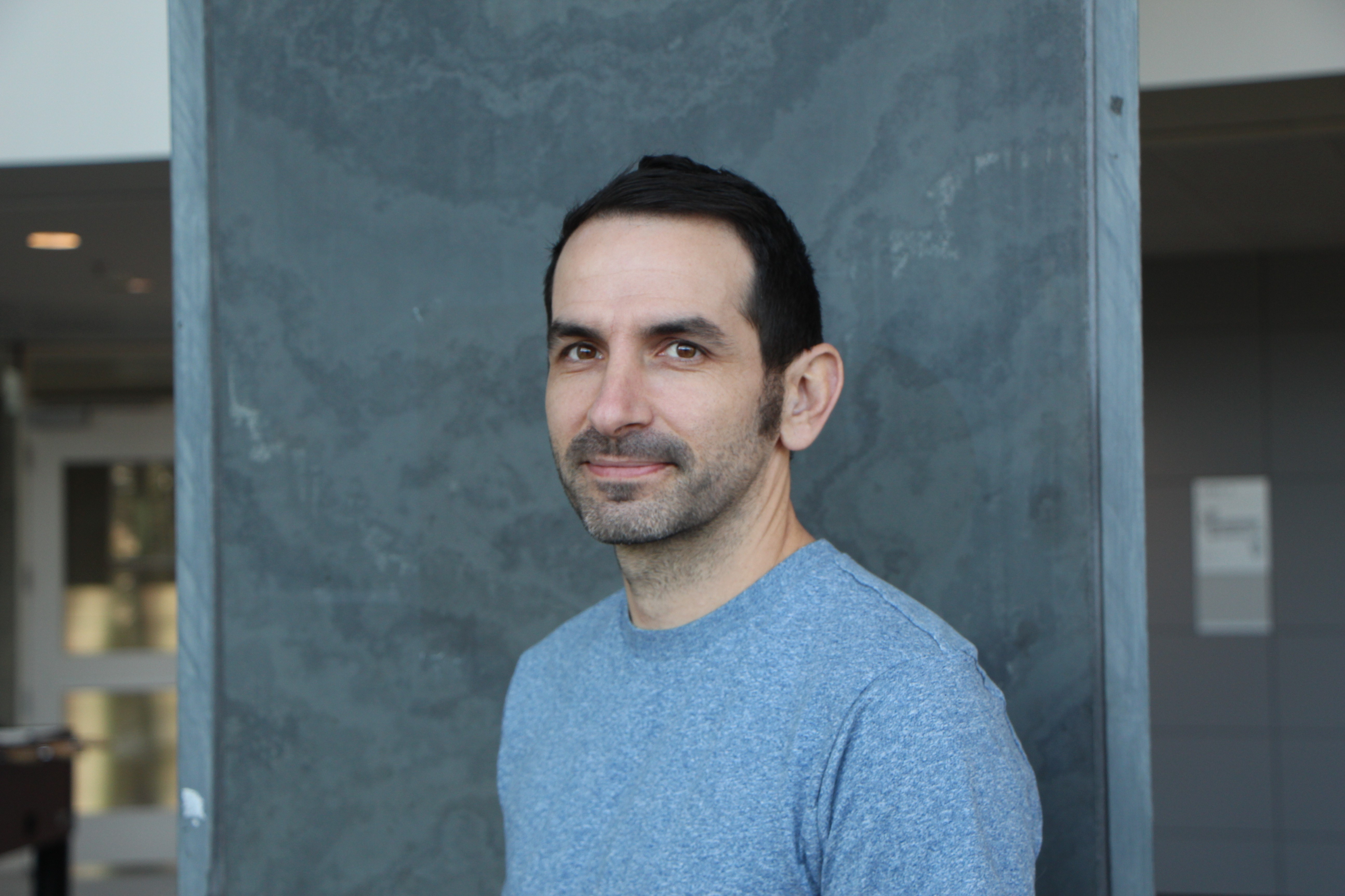
Dr. Chappleboim studies how cells communicate during a developmental process called somitogenesis, which drives the formation of repeated structures such as the spinal vertebrae. The signals that guide cell communication during this process can get misinterpreted by cancer cells, resulting in uncontrolled growth. These pathways are implicated in numerous cancer types but are notably associated with colorectal, ovarian, and breast cancer. Using cutting-edge techniques in human stem cells and 3D-models called organoids, along with the tools of computational biology, Dr. Chappleboim aims to deliberately perturb and examine these signaling pathways to gain a comprehensive understanding of how they function. Dr. Chappleboim received his PhD, MS, and BS from Hebrew University of Jerusalem, Jerusalem.
Fanglue Peng, PhD
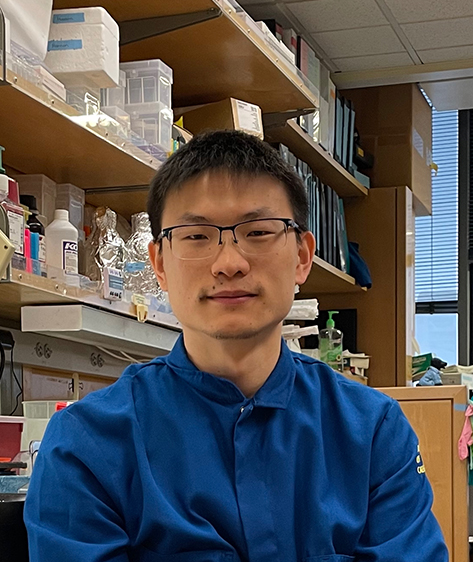
Accumulating evidence shows that specialized structures of white blood cells (lymphocytes), named tertiary lymphoid organs (TLOs), can form inside tumors and play a crucial role in fighting cancer progression. Unlocking the formation and functions of TLOs holds great promise for advancing cancer immunotherapy, but studying TLOs remains challenging due to the substantial disparities between humans and animal models. To address this, Dr. Peng [Connie and Bob Lurie Fellow] will leverage single-cell sequencing data and high-throughput screening methods to investigate a key initiator of TLO formation in human tumors. He further plans to develop innovative genetic models that enable the study of TLOs in a human-specific context within living organisms. By unraveling the intricacies of TLO biology, Dr. Peng aims to uncover novel therapies that can augment cancer immunotherapy and enhance treatment outcomes across various cancer types. Dr. Peng received his PhD from Baylor College of Medicine, Houston and his BS from Zhejiang University, Hangzhou, Zhejiang.
Ronghui Zhu, PhD
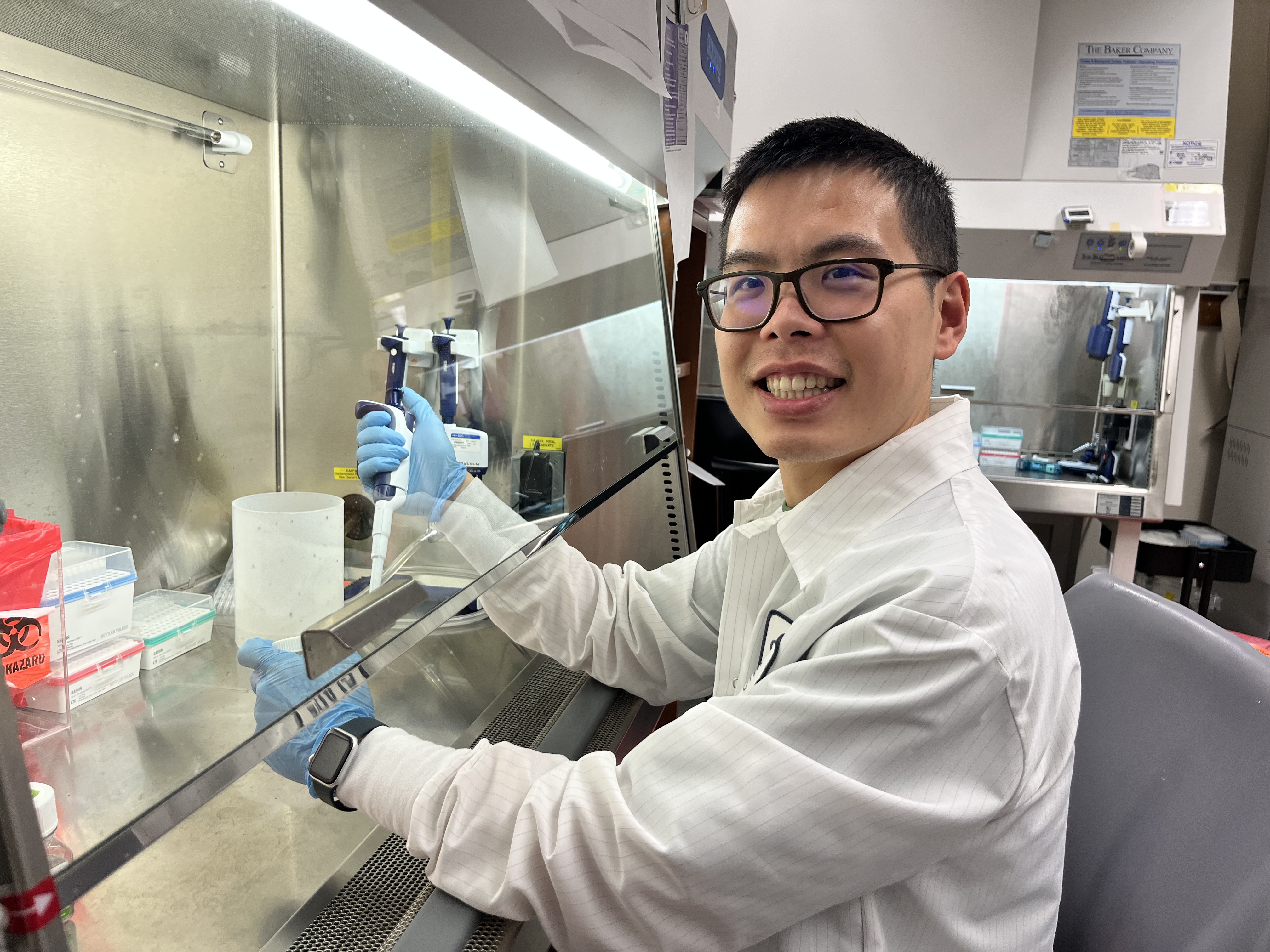
Our immune system can help us prevent or slow cancer development. Human CD4+ T cells play critical roles in regulating our immune responses to fight cancer. Upon encountering a pathogen, naïve CD4+ T cells differentiate into different T helper (Th) cells to perform diverse immune-modulatory functions. Variability in this differentiation process is associated with variable responses to cancer immunotherapy. While several genes necessary for differentiation have been identified, researchers lack a comprehensive map and a predictive model of the larger gene regulatory network (GRN) controlling this process. Dr. Zhu [Connie and Bob Lurie Fellow] plans to combine functional genomics with mathematical modeling to systematically map and model the human CD4+ T cell differentiation GRN and use the GRN model to predict and control the differentiation process. His work promises to provide a quantitative understanding of the CD4+ T cell differentiation process and open up new strategies for safer and more effective cell-based cancer therapy. Dr. Zhu received his PhD from the California Institute of Technology, Pasadena and his BS from Hong Kong University of Science and Technology, Hong Kong.
Isabella Fraschilla, PhD
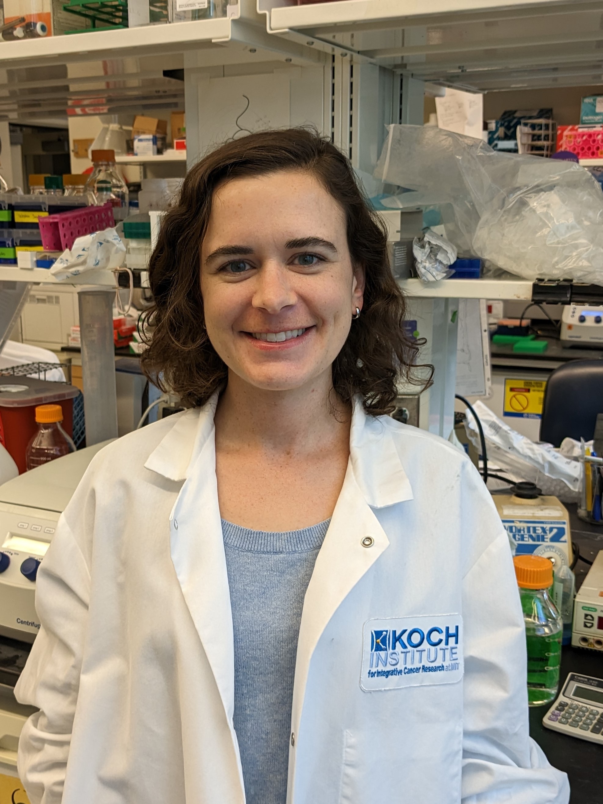
Pancreatic cancer remains unresponsive to current chemotherapy and immunotherapy treatments. However, with the recent development of mRNA vaccines and drugs that target cancer cell mutations, there is hope for a new generation of immune-based therapies. The ability of adaptive immune cells, called cytotoxic T cells, to kill cancer cells is central to anti-tumor immunity. Using mouse models of human pancreatic cancer, Dr. Fraschilla [Merck Fellow] plans to identify the flags presented by cancer cells that enable T cells to recognize them as foreign and kill them. One category of flags that label cancer cells as foreign may be proteins from bacteria that prefer to replicate within the tumor environment. This investigation of cancer cell targets will inform the development of future vaccines to treat cancer and prevent tumor regrowth or metastases. Dr. Fraschilla received her PhD from Harvard University, Cambridge and her BS from Emory University, Atlanta.
Wei (Will) Chen, PhD
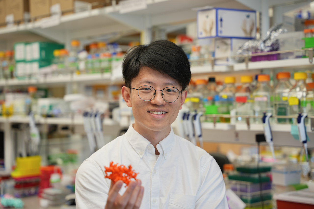
For gene activation, transcription factors (TFs) must bind to enhancers, often with multiple TFs binding at the same site, and recruit other proteins known as cofactors and polymerases. The interactions between TFs and cofactors are usually nonspecific, meaning the cofactors are interchangeable, which limits our understanding of precise gene activation. Dr. Chen will design new proteins that bind the cofactors with high specificity to clarify the contribution of each cofactor. This research will not only provide new insights into the mechanism of gene regulation but also provide new platforms to modulate gene expression with high precision. Dr. Chen received his PhD from the University of Washington, Seattle, his MS from Cornell University, Ithaca and his BS from Shandong University, Jinan.
Juner Zhang, PhD
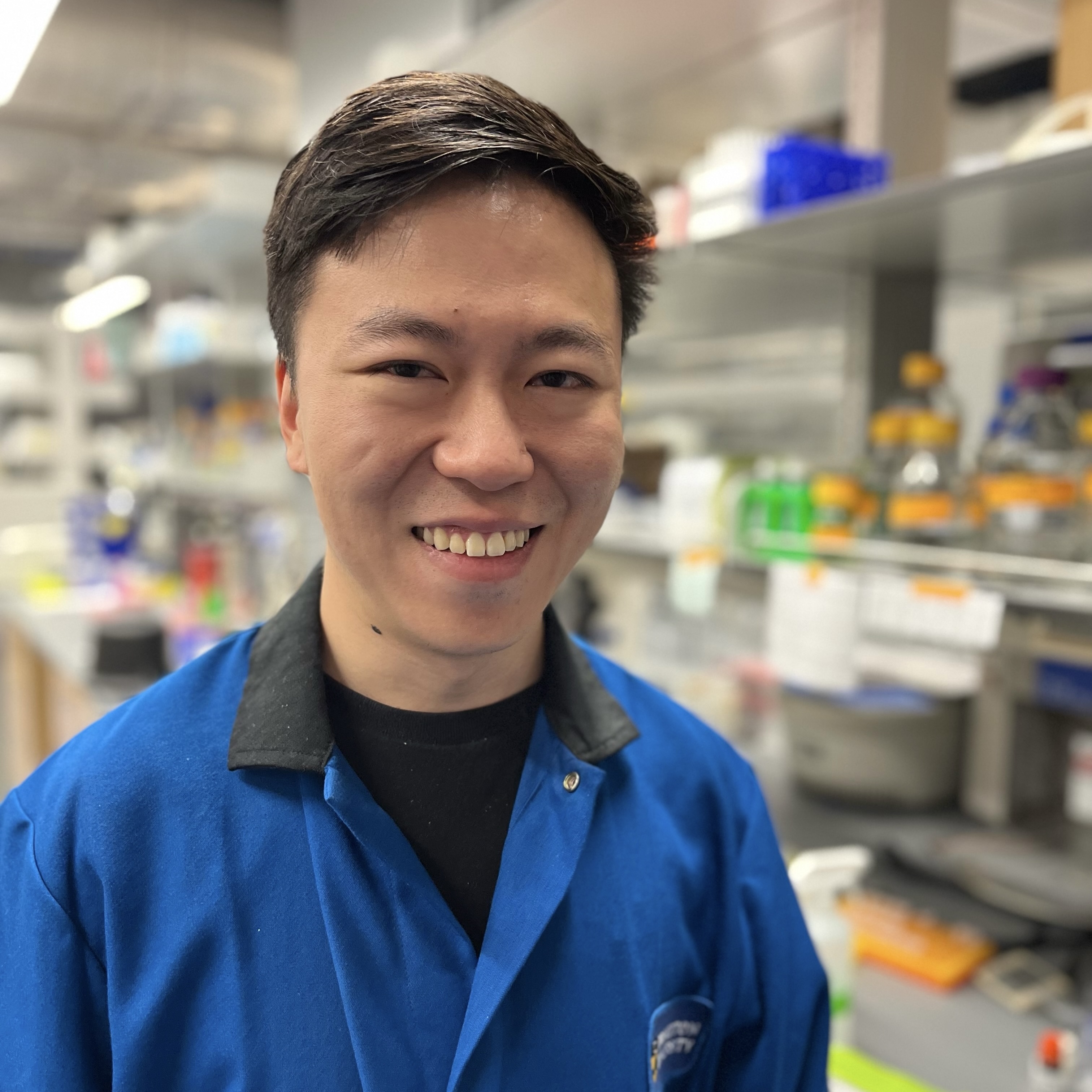
In cells, DNA wraps around a protein complex consisting of proteins called histones. Chemical modifications to histones can affect gene expression, which is key to activating or suppressing cancer progression. Histone monoaminylation, in which an amine (e.g., serotonin, dopamine, or histamine) attaches itself to a histone, is a newfound type of epigenetic modification whose role remains elusive in these processes. Dr. Zhang is using chemical biology tools to study the functions of these modifications as well as their effects on other adjacent, pre-existing cancer-associated modifications. This research may establish a foundation for how this epigenetic modification regulates gene expression and offer insight into the role of amines in the progression of cancer and human neurodegenerative disorders. Dr. Zhang received his PhD from the California Institute of Technology, Pasadena and his BS from Tsinghua University, Beijing.
Anders B. Dohlman, PhD
In many cancer types, microbiota have emerged as an influential component of the tumor environment. Dr. Dohlman [Meghan E. Raveis Fellow] studies Fusobacterium nucleatum, a bacterial species that colonizes around half of colorectal tumors. The reasons for F. nucleatum’s preferential colonization of these tissues are poorly understood, and investigating this phenomenon could lead to improvements in cancer diagnosis and treatment. To this end, Dr. Dohlman is using computational methods to study strains of cancer-associated F. nucleatum, searching for genomic features that promote colonization of colorectal cancers. In parallel, he is analyzing the genomes of colorectal tumors to identify genetic changes that in turn promote F. nucleatum colonization. Dr. Dohlman received his PhD from Duke University, Durham and his BA from Wesleyan University, Middletown.
Gabriel Cavin-Meza, PhD
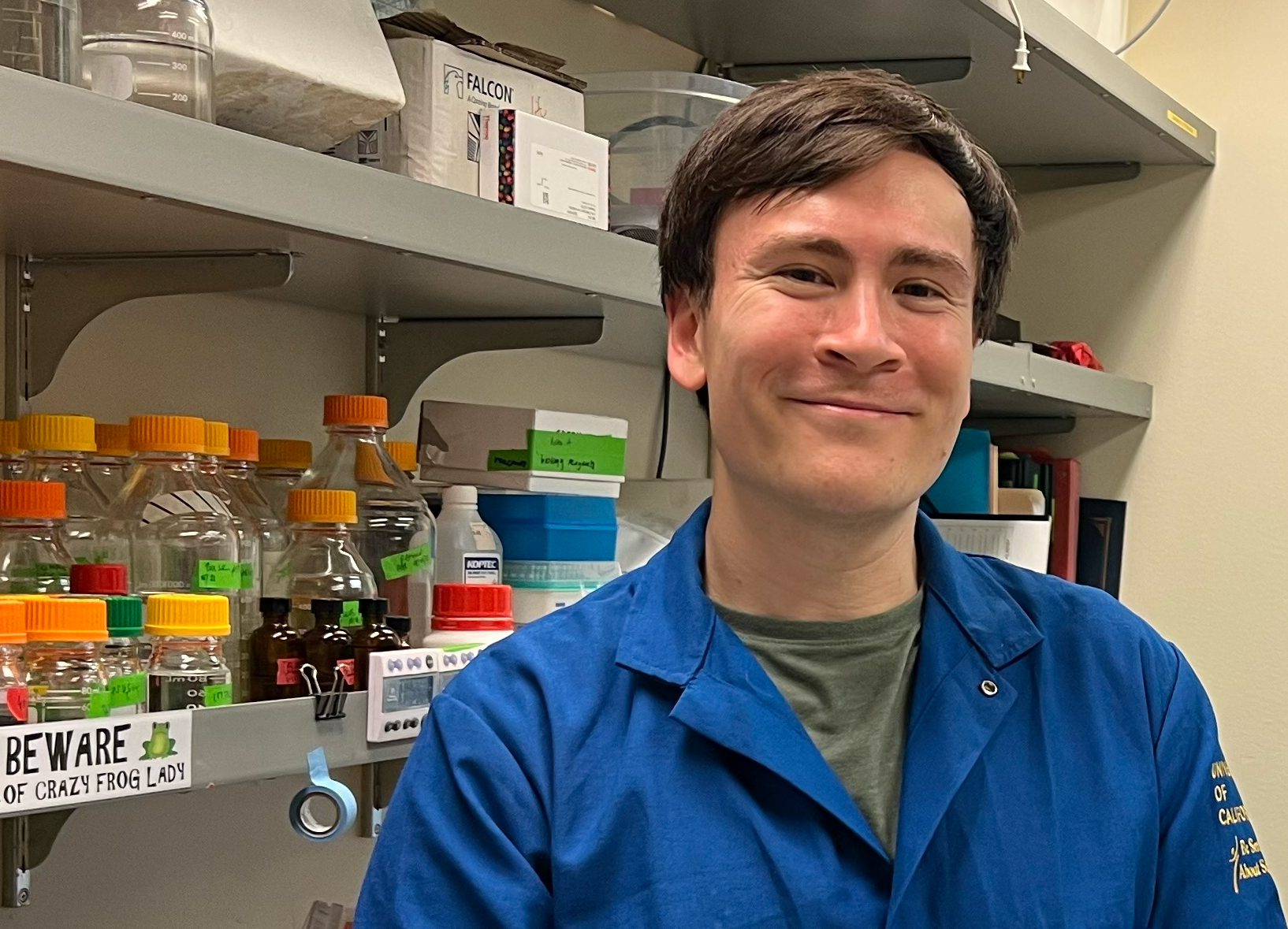
Proper cell division, including equal partitioning of DNA into two “daughter” cells, is critical for cell viability. However, many cancers continue to divide despite having atypical numbers of chromosomes and can even contain additional copies of the entire genome (polyploidy). Understanding how large increases in chromosome number affect cell division machinery has been limited by the methods used to generate polyploid cells. Serendipitously, stable polyploidy has arisen in multiple organisms, such as plants, fish, and amphibians. By utilizing the natural polyploidy found in Xenopus clawed frogs (ranging from two copies to twelve copies of the genome), Dr. Cavin-Meza [Merck Fellow] will explore the mechanisms that lead to increased but stable genome size. He will also analyze the proteome across Xenopus species to reveal how proteins have adapted to promote stable polyploidy over time, giving valuable insight into how stable polyploidy could arise in cancers. Dr. Cavin-Meza received his PhD from Northwestern University, Evanston and received his BS from the University of California, San Diego.
Pu Zheng, PhD
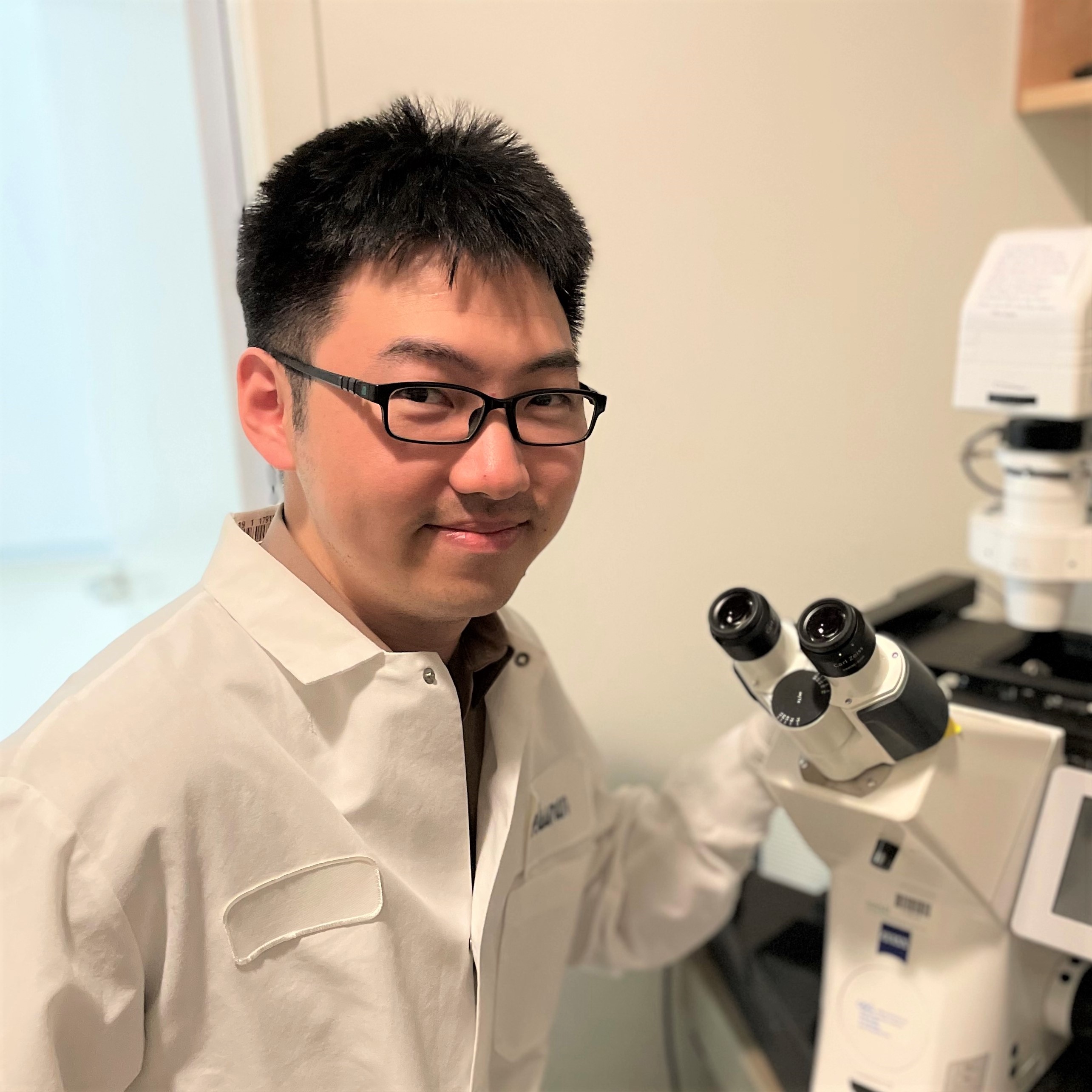
Dr. Zheng [Fayez Sarofim Fellow] is dedicated to the development of technologies for studying tumor evolution within their native contexts. Understanding the complex processes of cancer growth and progression requires a deep exploration of the dynamic interactions between tumor cells and the tumor microenvironment. “Spatial-omics” technologies are powerful tools that offer direct visualization of cells and their interactions in natural contexts, enabling systematic investigation of these intricate processes. Dr. Zheng aims to develop novel spatial-omics technologies that combine imaging and gene sequencing approaches to uncover the mechanisms underlying the spatially distinguished features of tumor evolution. Dr. Zheng received his PhD from Harvard University, Cambridge and his BS from Peking University, Beijing.
Nicole M. Hoitsma, PhD
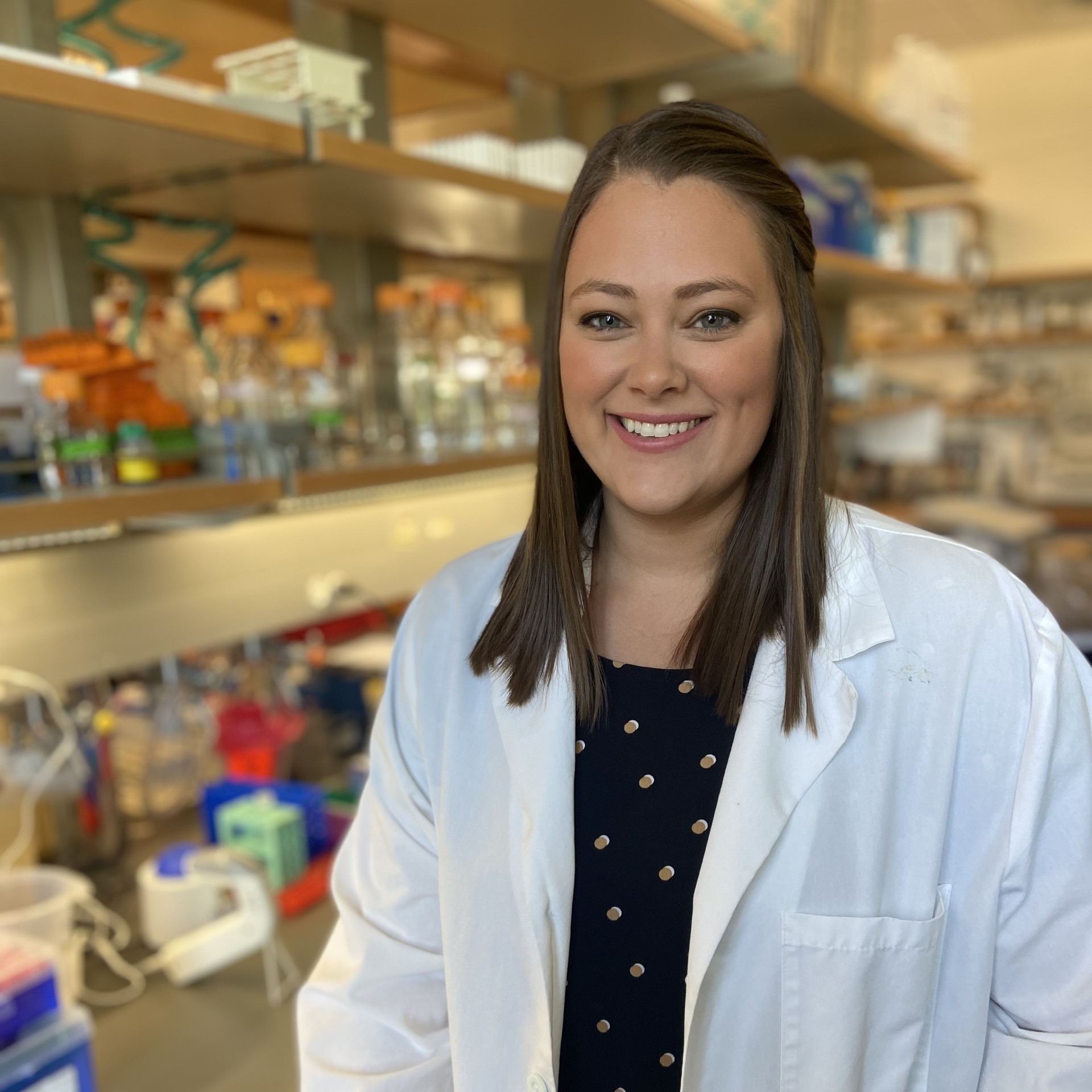
Human cells have complex mechanisms to repair DNA damage, such as that caused by exposure to sunlight or chemical substances. If DNA is not properly repaired, however, it can lead to cancer. In fact, faulty DNA repair has been associated with the initiation and progression of all types of cancer and is often targeted in cancer treatment to stop uncontrolled cell growth. A better understanding of how cells naturally defend against DNA damage will allow for the development of better drugs to treat cancer. Dr. Hoitsma [HHMI Fellow] aims to investigate specialized proteins, known as chromatin remodelers, that make damaged DNA accessible for repair. This research will provide insight for the development of novel therapeutic strategies to target these critical pathways. Dr. Hoitsma received her PhD from University of Kansas Medical Center, Kansas City and her BS from South Dakota State University, Brookings.
Nicholas P. Lesner, PhD
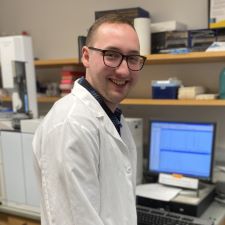
Ammonia, a waste product of cellular activity, is cleared from the body by the liver and kidneys through a process known as the urea cycle. During the urea cycle, ammonia is converted to urea, and arginine (an amino acid) is generated. When liver cells become cancerous, the urea cycle pathway stops functioning and cancer cells must import arginine from outside the cell. When cancer cells are prevented from importing arginine (via removal of arginine from the diet or genetic removal of the transporter), tumors do not grow, suggesting that arginine is critical for cells. However, the function of arginine in the cell is unclear. Using mass spectrometry and mathematical modeling, Dr. Lesner will identify the fate of arginine as it is metabolized by liver cancer cells in mouse models, and investigate how this is altered by various genetic mutations. Additionally, he will examine how restricting arginine from the diet genetically alters the liver and tumor cells. By understanding how disruption of this metabolic pathway influences liver cancer growth in the context of specific cancer drivers, Dr. Lesner aims to inform new therapeutic strategies. Dr. Lesner received his PhD from The University of Texas Southwestern Medical Center, Dallas and his BA from the University of Wooster, Wooster, Ohio.
Claudia A. Rivera Cifuentes, PhD
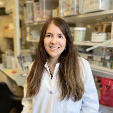
Endogenous retroviruses are viral elements of the human genome derived from retroviral infections of distant ancestors. Recent findings support the idea that these elements can cause immune system activation and inflammation. However, the crosstalk between endogenous retroviruses and the gut microbes that control immunity within the gut-and how abnormalities in this dialogue lead to inflammatory disorders-is not well understood. Further, although endogenous retroviruses have been proposed as potential targets for immunotherapy, we lack a mechanistic understanding of their interactions with the gut microbiota and how these interactions influence cancer development. Dr. Rivera Cifuentes [Lorraine W. Egan Fellow] aims to uncover how multi-kingdom interactions in the gut control intestinal health and colorectal cancer development. This work may have important clinical implications for the treatment of gut inflammatory disorders and gastrointestinal cancers. Dr. Rivera Cifuentes received her PhD from the University of Paris, Paris and her BSc from The Pontifical Catholic University of Chile, Santiago.
McLane Watson, PhD
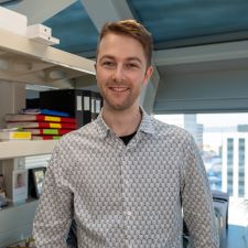
Cancer immunotherapy has revolutionized the way we treat cancer; however, it is only successful in a small subset of patients. Optimally functioning CD8 T cells, the specialized killers of the immune system, are key to the success of cancer immunotherapies. While CD8 T cell function is highly influenced by their metabolism, little is understood about how metabolism changes the function of these cells. Dr. Watson hypothesizes that metabolism affects CD8 T cell function by altering how tightly its DNA is packaged (its epigenetics), leading to altered gene expression. Using a mouse model of adoptive T cell therapy, a widely used immunotherapy in humans, and epigenetic techniques, Dr. Watson proposes to uncover how metabolism influences CD8 T cell epigenetic landscapes to control their function. He plans to apply these findings to improve T cell function and enhance tumor clearance. Dr. Watson received his PhD from the University of Pittsburgh, Pittsburgh and his BS from Hope College, Holland, Michigan.
Marie R. Siwicki, PhD
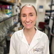
Neutrophils are important anti-microbial cells within the innate immune system. Recently, it has been shown that neutrophils can perform diverse functions, taking on both pro-inflammatory and pro-healing roles in response to tissue injury or insult. Dr. Siwicki's [Dale F. and Betty Ann Frey Fellow] goal is to understand how different neutrophil subtypes or states function to balance inflammatory versus regenerative processes, ultimately influencing tissue health and cancer. This work has the potential to uncover the basis of neutrophils' pro-tumor versus anti-tumor functions and could open the door to therapeutic targeting of specific neutrophil behaviors in order to improve clinical outcomes in cancer. Dr. Siwicki received her PhD from Harvard Medical School, Boston and ScB from Brown University, Providence.
James Swann, VetMB, DPhil
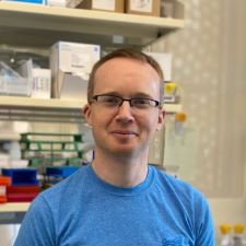
A key question in cancer biology is how genetic mutations, acquired over time, interact with environmental factors to affect emergence and progression of disease. This is particularly relevant in blood cancers because many people acquire genetic mutations in blood-forming stem cells in the bone marrow but only a small proportion go on to develop acute myeloid leukemia (AML). Dr. Swann [William Raveis Charitable Fund Fellow] is investigating whether inflammatory signals alter the behavior of stem cells that have already acquired an initial mutation, causing them to acquire features of cancer that will hasten the onset of AML. Specifically, Dr. Swann is interested in whether pre-cancerous stem cells change their gene expression in response to inflammation, which might allow them to outcompete normal cells in the bone marrow. He is utilizing cutting-edge techniques such as CRISPR editing of blood stem cells to investigate the molecular pathways responsible for these biological changes. This project has the potential to identify molecular pathways activated by inflammation that might promote AML development, offering new targets for therapeutic interventions. Dr. Swann received his VetMD (DVM) from the University of Cambridge and his DPhil (PhD) from the University of Oxford.
Mingjian Du, PhD
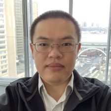
Global increases in metabolic syndrome, obesity, and diabetes are likely related to the overconsumption of hyper-palatable, cheap, ultra-processed food containing high amounts of added sugar and fat. Intriguingly, the vagus nerve has been discovered as the key conduit relaying information about sugar or fat ingestion from the gut to the brain, where a preference for sugar or fat is then developed and reinforced. Dr. Du [HHMI Fellow] aims to understand how the neurons are organized in the gut-brain vagal axis to sense sugar and fat, and to identify and characterize the neural circuits downstream of the gut-brain vagal axis that produce an insatiable appetite for sugar and fat. Understanding the basic biology of the gut-brain axis can provide important insights and strategies to help combat overconsumption of highly processed foods rich in sugar and fat, which may contribute to lowering the risk of metabolic diseases and cancer. Dr. Du received his PhD from The University of Texas Southwestern Medical Center, Dallas and his BS from the Tsinghua University, Beijing.
Christopher Noetzel, PhD
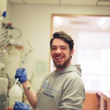
As different tissues in the body form, cells need to undergo a complex, precisely timed series of differentiation programs to form specialized cell types. Importantly, premature or delayed initiation of these programs can contribute to cancer formation. However, how timing of cellular differentiation is encoded on a molecular level is poorly understood. Dr. Noetzel is using the protozoan parasite Cryptosporidium parvum as a simplified model of eukaryotic differentiation. After infecting the intestinal lining of a mammalian host, these single-celled parasites undergo exactly three rounds of asexual replication before collectively differentiating into gametes. These studies will investigate how this hard-wired, intrinsic developmental timer is encoded. In his project, Dr. Noetzel aims to understand how these parasites "count to three," which will inform our basic understanding of how eukaryotic cells keep track of time during development. Dr. Noetzel received his PhD from the Weill Cornell Medical College, Cornell University, New York and his MSc and BSc from Georg-August-University, Göttingen.
Archana Krishnamoorthy, PhD

Cancer initiation and progression stems from cell division errors that promote chromosome breakage and accumulation of mutations. Dr. Krishnamoorthy [HHMI Fellow] will use cutting-edge, cross-disciplinary approaches to provide insights into the fundamental question of how cell division shapes the cancer genome. Understanding the mechanisms of cancer genome complexity will help identify better diagnostics and treatments for cancers linked with high levels of genome alterations. Dr. Krishnamoorthy received her PhD from Vanderbilt University, Nashville and her MS from Middle Tennessee State University, Murfreesboro and her BS from PES Institute of Technology, Bangalore.
Joshua B. Sheetz, PhD
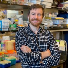
Cancer cells adapt their metabolism to achieve rapid growth and proliferation. Much of their metabolic malleability hinges on mitochondria, subcellular hubs for energy transformation and biosynthesis. As a key means to control mitochondrial composition and meet metabolic demands, cells mark mitochondrial proteins for degradation by a process called ubiquitylation. How both cancerous and healthy cells direct and monitor mitochondrial ubiquitylation remains poorly understood. Dr. Sheetz [HHMI Fellow] aims to dissect the cellular machinery that performs mitochondrial ubiquitylation and determine how this process promotes metabolic adaptability in cancer cells. A major translational goal is to identify approaches for tuning the levels of mitochondrial ubiquitylation in tumors and in metabolic disorders that put patients at risk for cancer. Dr. Sheetz received his PhD from Yale University, New Haven and his BS from the University of North Carolina, Chapel Hill.
Ben F. Brian, PhD
Abnormal interactions between our immune system and our gut microbes can lead to inflammation that drives colon and gastric cancer growth. Dr. Brian [HHMI Fellow] is investigating how the immune system recognizes and responds to these microbes, and how these interactions contribute to abnormal inflammation that can fuel cancer growth. Microbiota-immune interactions have been generally studied in the context of "clean" laboratory mice, but these models do not fully capture human immunology and the complex interplay between host cells and foreign microbes. To overcome this, Dr. Brian plans to study these interactions in "dirty" mice, colonized by a diverse community of microbes as well as pathogens. He will then use laboratory mice with more defined microbial communities to test how recognition of specific microbes by the immune system is regulated and how disruptions to this regulation contributes to inflammation. Dr. Brian received his PhD from the University of Minnesota, Twin Cities and his BS from the University of California, Santa Barbara.
Ryan Y. Muller, PhD
The PABPC1 protein has diverse roles in gene expression control that span functions in mRNA stability, polyA tail length control, and translation regulation. PABPC1 gene amplifications are detected in roughly 4% of cancer samples, but it is unclear how PABPC1 fits into the picture of cancer progression. Dr. Muller [HHMI Fellow] studies the sequence preferences of PABPC1 protein to understand the mechanistic details that determine which transcripts are subject to PABPC1-mediated regulation. Connecting these sequence preferences to the mis-regulation caused by excess PABPC1 may provide a therapeutic handle for cancers that contain PABPC1 gene amplifications. Dr. Muller received his PhD from the University of California, Berkeley and his BS from Arizona State University, Tempe.
Hui (Vivian) Chiu, PhD
Fatigue is the most common symptom experienced by patients with cancer or undergoing cancer treatment. While chronic inflammation and hormonal imbalance have been suggested as possible causes, the roots of cancer-related fatigue remain unclear and thus we lack effective treatments. Dr. Chiu [HHMI Fellow] seeks to illuminate the physiological basis of fatigue using interdisciplinary approaches that combine the strengths of neuroscience, immunology, and computational biology. Through the lens of brain-body interactions, Dr. Chiu aims to identify key molecular and cellular components of fatigue with the goal of improving treatments for cancer and other severe diseases, such as long COVID. Dr. Chiu received her PhD from the California Institute of Technology, Pasadena and her MS and BS from the National Taiwan University, Taiwan.
Erik Van Dis, PhD
The innate immune system is the body's first line of defense against pathogens. The innate immune sensor MDA5 detects nucleic acids derived from pathogenic genomes or damaged cells and drives the production of cytokines, an important signaling molecule in the immune inflammatory response. MDA5 can be aberrantly activated by host nucleic acids, however, leading to autoimmune activation. Hyperactive MDA5 alleles are associated with the development of autoimmune diabetes. Dr. Van Dis [Robert Black Fellow] aims to define the innate immune signaling pathways that initiate autoimmune diabetes to better understand immune activation pathways in the pancreas and guide the development of novel immunotherapies for pancreatic cancer. Dr. Van Dis received his PhD from the University of California, Berkeley and his BA from Carleton College, Northfield.
Tadashi Manabe, MD, PhD
Lung cancer remains the leading cause of cancer mortality. Substantial breakthrough discoveries, including the identification of lung cancer-specific genetic drivers (e.g., EGFR mutations, EML4-ALK fusion genes) and the development of molecular inhibitors of these pathogenic factors, have improved outcomes for patients with advanced-stage lung cancer. However, lung cancer cells eventually acquire resistance to these molecular inhibitors, resulting in progressive disease. Dr. Manabe’s [Connie and Bob Lurie Fellow] research focuses on protein compounds formed by the self-assembly of oncogenic fusion proteins such as EML4-ALK. These compounds initiate a signaling pathway that causes abnormal cell proliferation in cancer. Dr. Manabe will explore the newly discovered structures of signaling proteins with the goal of developing molecular therapies that enhance precision medicine strategies and improve the control of lung cancer. Dr. Manabe received both his MD and PhD from Keio University School of Medicine.
Titas Sengupta, PhD
An organism’s life experiences, such as exposure to bacterial pathogens, can cause sustained changes in its physiology and behavior. How these experiences are encoded in heritable RNA and DNA-associated proteins (called chromatin), and how these in turn affect the physiology of the organism itself and its progeny, are not well understood. Previous research has shown that the roundworm C. elegans can “read” small non-coding RNAs from the pathogenic bacterium Pseudomonas aeruginosa and learn and teach its progeny to avoid this bacterium. Dr. Sengupta’s [Rebecca Ridley Kry Fellow] research investigates how bacterial small RNAs taken up in the intestine can result in lifelong, multigenerational, and organism-wide changes at the epigenetic (RNA and chromatin) level to regulate brain function and behavior. She will investigate which small RNA and chromatin-associated genes are required for the learned response, where these genes function, and what changes at the epigenetic and gene expression level underlie this response. This will inform principles of epigenetic regulation of gene expression following diverse environmental stimuli, and stimuli within tissue environments, including tumor microenvironments. Dr. Sengupta received her PhD from Yale University and her MS and BS from the Indian Institute of Science Education and Research.
Manuel Osorio Valeriano, PhD
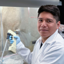
Human cells compact their vast genomes into the small confines of the nucleus by wrapping their DNA into a highly complex structure called chromatin. Packaging DNA into chromatin, however, affects all nucleic acid-transacting machines (e.g., transcription factors) that need to access the genomic information stored in the DNA. NuRD is a large multi-subunit protein complex that plays a major role in making chromatin either accessible or inaccessible. Dysregulation of NuRD and aberrant targeting of the complex can result in the emergence of several types of cancers, including breast, liver, lung, blood, and prostate cancers. Dr. Osorio Valeriano’s [Philip O'Bryan Montgomery, Jr., MD, Fellow] work will reveal mechanistic aspects of NuRD-mediated chromatin regulation and pave the way for the development of novel therapeutic approaches that target cancers more effectively. Dr. Osorio Valeriano received his PhD from Philipps University and his MSc and BSc from the National Autonomous University of Mexico.
Senén D. Mendoza, PhD
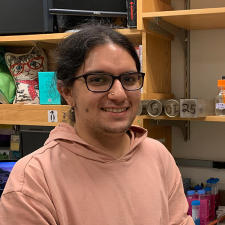
In addition to acute illness, viruses can cause cancers. While our understanding of cellular immunity against viruses that have DNA-based genomes is robust, we know less about how cells protect themselves against RNA-based viruses such as hepatitis C, a leading cause of liver cancer. Because many cellular defenses against viruses are known to be shared between mammals and bacteria, Dr. Mendoza [HHMI Fellow] is looking for new cellular defenses against RNA viruses in bacteria and will investigate how these defenses work. The resulting discovery of anti-viral defenses will broaden our understanding of how cells protect themselves against RNA viruses, which will improve our capacity to support patients' immune systems when infected with cancer-causing RNA viruses. Dr. Mendoza received their PhD from the University of California, San Francisco, and their BS from the University of Miami.
Wen Mai Wong, PhD
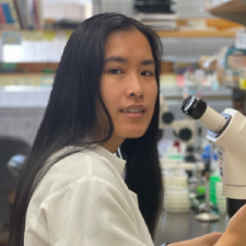
Multiple cancers, including prostate, breast, and gastrointestinal cancers, are known to be heavily innervated. However, the role of neurons and their signaling within the tumor microenvironment remains unknown. Previous work has shown that transecting the vagus nerve can block the progression of gastric cancer, emphasizing a critical role for the vagal neurons in this disease. However, these transections produce side effects, making it a difficult strategy to translate to the clinic. Dr. Wong [Kenneth G. and Elaine A. Langone Fellow] is proposing a new method to non-invasively silence neurons within the body. Specifically, she will use ultrasound to silence specific neurons in rodent models in order to determine the impact of these neurons on animal behavior and disease physiology, including the tumor microenvironment. Dr. Wong received her PhD from the University of Texas Southwestern Medical Center and her BS from St. Mary’s University.
Rebecca S. Moore, PhD

Sleep problems may be a risk factor for developing certain types of cancer—lung, colon, pancreas, and breast—and may affect the progression of these cancers and the effectiveness of their treatment. Conversely, symptoms of cancer or side effects of treatment, including restless legs and obstructive sleep apnea, may cause sleeping problems, reducing quality of life. Understanding the complex relationship between cancer and sleep creates opportunities to improve health, treatment options, and quality of life. Specifically, understanding how the peripheral nervous system and the brain regulate both the timing and rhythmicity of sleep (i.e., circadian control), and the balance between time awake and growing sleep pressure (i.e., homeostatic control), could improve survival rates and the quality of cancer treatment. To this end, Dr. Moore [HHMI Fellow] aims to identify the role of circulating dietary cholesterol on sleep and to conduct a targeted genetic screen to identify peripherally secreted proteins that affect either the circadian or the homeostatic control of sleep. These results will provide a means for therapeutic interventions to ameliorate the effects of sleep disruption. Dr. Moore received her PhD from Princeton University and her MS and BS from the City College of New York.
Erron W. Titus, MD, PhD
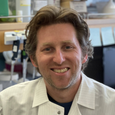
Chimeric antigen receptor (CAR) T cells are immune cells that have been genetically engineered to bind specific proteins on cancer cells. CARs can display exquisite sensitivity and discrimination, and CAR T cells have been deployed with spectacular success to detect and kill blood cancers. Unfortunately, they are much less effective against “solid” tumors, such as breast or kidney cancers. To address this problem, Dr. Titus [Connie and Bob Lurie Fellow] is designing T cells with membrane proteins that perform novel functions, including proteins that facilitate membrane fusion or alter the adhesion between T cells and their targets. By redesigning T cell membranes, Dr. Titus hopes to create useful cancer-fighting tools that can be deployed in conjunction with other emerging cellular therapies and immunotherapies. Dr. Titus received his MD and PhD from the University of California, San Francisco, and his AB from Harvard University.
Dylan M. Parker, PhD
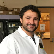
Dr. Parker [HHMI Fellow] studies the role of molecular assemblies known as stress granules that form when cells are exposed to stressful conditions. The assembly of stress granules upon cellular insult is thought to regulate gene expression and modulate cell survival. Notably, stress granules are present in various cancers and many chemotherapeutic treatments lead to the formation of stress granules. Dr. Parker aims to determine the mechanisms regulating stress granule assembly and disassembly to understand how stress granule formation supports the development of cancer and chemotherapy-resistant tumors. This research has the potential to discover novel targets to treat cancers as well as sensitize chemotherapy-resistant cancers to existing treatments. Dr. Parker received his PhD from Colorado State University and his BS from the University of Oregon.
J. Scott P. McCain, PhD
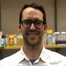
One of the defining features of cancerous cells is that they divide quickly. The composition of the human microbiome is also due to differences in how quickly microbes grow. How do we determine how fast cells are growing in their natural environment? Is there a way to take a ‘snapshot’ and turn it into a ‘growth rate’? This is the fundamental problem Dr. McCain is studying. He is using computational simulations, machine learning, and experiments with bacteria to determine the optimal way to use markers of gene expression to estimate these critical rates. This project will provide fundamental insights into the use of gene expression data to key processes like growth rate or metabolite secretion rate, both of which have implications for cancer biology. Dr. McCain received his MSc and PhD from Dalhousie University and his BSc from the University of Western Ontario.
Grace E. Johnson, PhD
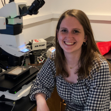
Dr. Johnson [HHMI Fellow] studies the role that a particular type of cell-cell communication, known as quorum sensing, plays in the development of spatially structured bacterial communities called biofilms. Biofilm formation promotes disease in many clinically relevant bacterial species, and infections caused by them pose severe risks for patients receiving chemotherapy. Dr. Johnson is currently investigating how quorum sensing within biofilms establishes patterns of gene expression, and in turn, how these patterns drive biofilm development and dictate biofilm architectural features. By defining mechanisms underlying biofilm formation and biofilm architecture, Dr. Johnson hopes to contribute to the generation of new approaches for disrupting quorum-sensing-controlled bacterial community interactions as a means of combating bacterial pathogens. Dr. Johnson received her PhD from MIT and her BS from Yale University.
Catherine Triandafillou, PhD
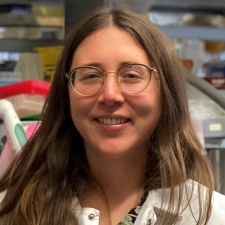
When an organism is developing, it must correct mistakes that might occur at the level of individual cells or tissues. Dr. Triandafillou [National Mah Jongg League Fellow] wants to better understand how error correction systems work, and why they might not work in cases like cancer. To explore these developmental questions, Dr. Triandafillou uses what are called gastruloids, 3D clusters of stem cells that can organize themselves and transform into the basic building blocks of an organism. She developed a method using microscopy to trace the history of these cells and measure how much their past state and history influence what they become. Dr. Triandafillou wants to see how differences in individual cells might impact what those cells eventually turn into, and how such differences affect the correction of mistakes like abnormal growth, bias in cell types, or missing cell types. She is also interested in how the cells around an error react to it. Dr. Triandafillou received her PhD from the University of Chicago and her BS from Temple University.
Zeda Zhang, PhD
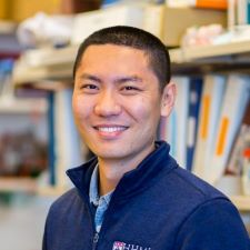
On the cellular level, aging manifests as cellular senescence—when cells permanently stop multiplying but do not die. Aberrant accumulation of senescent cells is thought to be a major contributor to age-dependent tissue degeneration and its associated pathologies. Elimination of senescent cells has been shown to improve age-associated tissue damage pathologies and extend healthy lifespan in mice. Senescent cells undergo extensive remodeling on their surface, including increased production of many surface proteins. Dr. Zhang [HHMI Fellow] is using a quantitative proteomics approach to investigate the mechanisms and biological consequences of cell surface remodeling in senescent cells. His goal is to identify new therapeutic targets on the senescent cell surface and develop next-generation chimeric antigen receptor (CAR) T cells and antibodies to evaluate their impact on age-related diseases. Success with this approach may have a transformative impact on treating life-threatening diseases like cancer, fibrosis, and atherosclerosis. Dr. Zhang received his PhD from Gerstner Sloan Kettering Graduate School and his BS from Sun Yat-Sen University.
Patrick Woida, PhD

Dr. Woida studies the foodborne pathogens Listeria monocytogenes and Shigella flexneri that enter and replicate within human cells. These bacteria also directly infect neighboring cells by pushing against the host cell membrane to form long membrane protrusions that extend and eventually release the bacteria into the new cell. This process of cell-to-cell spread requires the bacteria to hijack intercellular signaling pathways to reshape the host cell membrane. These signaling pathways normally regulate human cell adhesion and motility, and their dysregulation promotes tumor growth and metastasis. Dr. Woida’s goal is to uncover the unique mechanisms by which these pathogens remodel the host cell membrane to gain insight into how the co-opted intercellular signaling pathways function under both healthy conditions and tumor progression. Dr. Woida received his PhD from Northwestern University and his BS from the University of Illinois at Urbana-Champaign.
Siqi Li, PhD
Dr. Li [The Mark Foundation for Cancer Research Fellow] studies signaling events regulating the competition between cells carrying cancer-causing mutations and normal cells during cancer initiation. Previous studies have shown that intercellular signaling between mutant and normal cells could regulate the proliferation of these cells and shape the outcome of cancer initiation. Dr. Li is adapting novel tools to identify what molecular cues are mediating this crosstalk and how they contribute to cancer growth in mouse skin. Understanding these events may guide the development of cancer prevention strategies that restrict the early expansion of mutant cell lines in skin and other tissues. Dr. Li received her PhD from Duke University and her BS from Tsinghua University.
Henry R. Kilgore, PhD
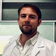
Cells are compartmentalized into membrane-bound and membrane-less organelles, providing spatial structure to the cell’s concentration of proteins and nucleic acids. Dr. Kilgore’s research aims to understand the environment inside different organelles and apply this knowledge to the development of targeted cancer therapies, as better targeting within the cell will improve drug efficacy, increase potency, and decrease side effects. Using both live cells and reductionist models, he will investigate how molecules distribute themselves within the cell as a function of their chemical properties. Learning and applying the chemical grammar of this spatial partitioning will enable the design and preparation of molecular probes and drugs that synergize with the chemistry of the cell as a mechanism of treating all cancers. Dr. Kilgore received his PhD from Massachusetts Institute of Technology and his BS from the University of California, Berkeley.
Elizabeth R. Hughes, PhD
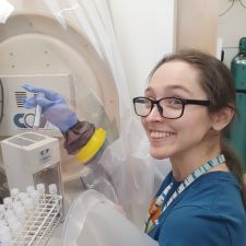
Immune checkpoint inhibitors, a type of cancer treatment that helps immune cells identify and kill tumor cells, have been a major breakthrough in the treatment of many cancer types. Unfortunately, not all patients respond to this immunotherapy. Dr. Hughes [Robert Black Fellow] is studying how gut microbes improve response to immune checkpoint inhibitors. The bacterium Akkermansia muciniphila lives in the gastrointestinal tract and has been shown to improve response to immune checkpoint inhibitors via poorly understood mechanisms. Dr. Hughes aims to discover how A. muciniphila improves response to cancer immunotherapies and to design microbe-based therapeutic strategies that will further enhance cancer immunotherapy responses. Dr Hughes received her PhD from UT Southwestern Medical Center and her BS from Baylor University.
Edward M. C. Courvan, PhD
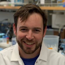
Macrophages are specialized immune cells responsible for “eating” harmful cells, presenting antigens to T cells, and initiating inflammation by releasing signaling molecules called cytokines. Macrophages could potentially be activated to attack tumor cells, but for reasons that are currently unclear, they instead signal for the tumor to grow faster and become more invasive. Dr. Courvan [HHMI Fellow] is investigating how macrophages respond to the low-oxygen environment inside tumors, and specifically how they regulate gene expression through post-transcriptional mechanisms in low-oxygen conditions. With this research, he hopes to uncover new ways to leverage the body's immune system against cancerous cells. Dr. Courvan received his PhD from Yale University and his BS from the University of Connecticut.
Fangtao Chi, PhD
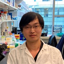
Dietary interventions such as caloric restriction (CR) and ketogenic diet (KD) are reported to limit tumor growth partially by modulating stem cell function. The intestine functions as the main organ of nutrient absorption and, due to rapid tissue renewal via intestinal stem cells (ISCs), is sensitive to shifts in the body’s metabolic state before and after eating. Both CR and KD conditions dramatically enhance the activity of an enzyme in ISCs known as HMGCS2. This enzyme controls ketogenesis, the conversion of fatty acids into ketone bodies as a means of producing energy when glucose is unavailable. Dr. Chi aims to dissect the role of ketone body metabolites in modulating intestinal stem cell function and tumor growth. With a better understanding of how intestinal stem cells adapt to diverse diets, he hopes to identify new strategies or dietary interventions that prevent and reduce the growth of cancers in the intestinal tract. Dr. Chi received his PhD from the University of California, Los Angeles and his BS from Zhejiang University.
Felix C. Boos, PhD
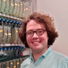
Evidence that aging is driven by defined, regulated processes (rather than simple “wear and tear”) has sparked hope that we might target these processes to fight age-related diseases. A particularly exciting example is the regulation of protein homeostasis, or the balance between protein synthesis, folding, and degradation. Protein homeostasis is deregulated in both cancer and normal aging, but the underlying mechanisms remain elusive. Dr. Boos will use the short-lived African turquoise killifish as a new model organism to study how different cells and tissues respond to protein misfolding, how they coordinate their responses, and how aging influences these pathways. This research will not only unravel fundamental mechanisms of aging, but also inform new strategies to fight multiple types of cancer. Dr. Boos received his PhD and his B.Ed. from the University of Kaiserslautern.
Debadrita Bhattacharya, PhD
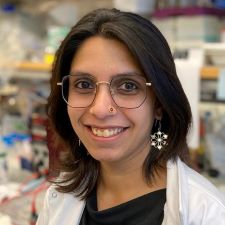
Intra-tumoral heterogeneity (ITH), or the evolution of distinct cell types within a tumor, underlies most fatal features of cancer and presents a great therapeutic challenge. Using small cell lung cancer (SCLC), a highly heterogeneous and lethal form of lung cancer, as a model, Dr. Bhattacharya [Robert Black Fellow] will study how ITH arises during cancer progression. She will employ emerging genomics techniques to characterize the cellular subtypes that comprise SCLC tumors and identify “druggable” transcription factors which, if targeted, could reduce tumor heterogeneity in this cancer. By profiling thousands of cells from treatment-naïve and therapy-resistant tumors, Dr. Bhattacharya aims to identify the “master-regulators” of the cellular subtypes that expand upon treatment in SCLC. She will then evaluate the role of these factors in human patient-derived cell lines, with the goal of uncovering novel mechanisms underlying ITH in human cancers. Dr. Bhattacharya received her PhD from Cornell University and her BS from the University of Calcutta.
Rico C. Ardy, PhD
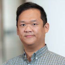
Dr. Ardy [Robert Black Fellow] is investigating the genetic determinants that govern the behavior of fibroblasts, a type of connective tissue cell that has been implicated in arthritis, heart disease, and cancer. Activated fibroblasts can exacerbate disease through various mechanisms, including remodeling tissue architecture and modulating the immune system. Dr. Ardy plans on using state-of-the-art genetic tools, including CRISPR inhibition and activation coupled with single-cell RNA sequencing technology, to uncover the proteins and pathways that regulate fibroblast behavior and thereby inform the development of new targeted cancer treatments. Dr. Ardy received his PhD from the Medical University of Vienna and his BS from the University of California, Los Angeles.
Charles H. Adelmann, PhD
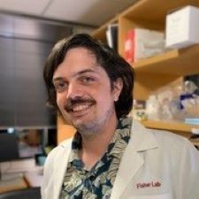
Skin cancer is the most common type of cancer worldwide, and sun exposure is known to be one of the main risk factors for developing skin cancers. Melanin pigment gives our hair, eyes, and skin their color, and it also shields skin cells from the carcinogenic effects of sun exposure. Combining just one enzyme (tyrosinase) and two substrates (oxygen and tyrosine) in the lab results in the generation of melanin—yet we know that dozens of other proteins affect pigmentation in humans. How does a process that requires so few components in vitro utilize these other factors in the human body? Dr. Adelmann’s work focuses on the cellular and biochemical contributors to human pigmentation, a clearer understanding of which will facilitate chemopreventative interventions for skin cancer that manipulate or mimic the anti-cancer properties of pigmentation. Dr. Adelmann received his PhD from Massachusetts Institute of Technology and his BA from Rice University.
Akanksha Thawani, PhD
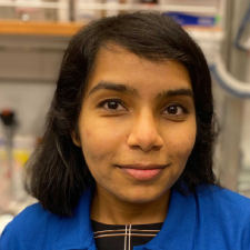
Dr. Thawani [Merck Fellow] studies selfish DNA sequences—so called because they copy and paste themselves within the human genome despite offering no specific fitness advantage. Dr. Thawani will utilize advanced methods such as cryo-electron microscopy to reveal the cellular machinery that assists these selfish elements and thus delineate their mechanism of mobility. She will use this insight to engineer new genome editing technologies to precisely insert large genes at user-specified sites in a variety of human cell types. This general technology will not only translate directly into new gene therapies, but also result in wide-ranging applications in synthetic biology. Ultimately, this work will contribute to treatment for many cancer types, including improved CAR-T therapies for blood cancers.
Ching-Ho Chang, PhD
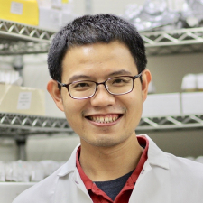
Dr. Chang is studying protamines—short, positively-charged proteins that condense DNA into chromatin and regulate gene expression in sperm nuclei. While eukaryotic cells use histones to package genomes in a way that allows access for transcription and replication, sperm cells must package their genomes more tightly. For this, many animals deploy protamines instead of histones. Despite sharing certain functions with highly conserved histones, protamines have independently arisen in evolution multiple times and are continuing to rapidly evolution. Using Drosophila fruit fly species as a model, Dr. Chang studies how sperm chromatin regulates gene expression and reproductive fitness. Additionally, although protamine expression is typically limited to testes, their misexpression has been observed in many cancers, indicating an opportunity for therapeutic intervention.
Qinheng Zheng, PhD
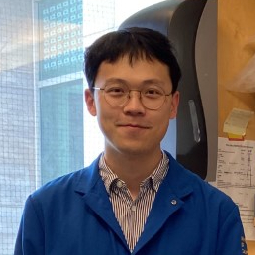
Dr. Zheng [Connie and Bob Lurie Fellow] is developing small molecules that selectively inhibit the protein K-Ras(G12D). Pancreatic ductal adenocarcinoma (PDAC) is the most lethal common cancer due to the infrequency of early diagnosis and the lack of targeted or immune therapies. A high percentage (>90%) of PDAC patients harbor KRAS mutations, with the majority expressing the K-Ras(G12D) missense mutation. Despite extensive drug discovery efforts across academia and industry, there are no approved drugs directly targeting oncogenic K-Ras(G12D). K-Ras lacks an apparent surface topology for reversible small molecule binding, leading to its notorious characterization as “undruggable.” Dr. Zheng is searching for small molecules that form a permanent bond with the mutant protein at its missense site and inhibit its interaction with effector proteins.
Jung-Shen Benny Tai, PhD
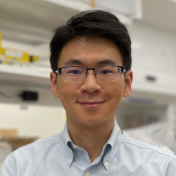
Dr. Tai studies bacterial biofilms or aggregates of bacterial cells in an extracellular matrix. Biofilms play a critical role in many health and industry settings. Biofilm-forming bacteria and imbalance in patients’ gut microbiota have been found to correlate with cancer development, and cancer patients receiving therapy frequently suffer from bacterial infections. From the unique perspectives of microbiology, soft matter physics, and ecology, Dr. Tai aims to decipher how, at the single bacteria cell level, heterogeneities in cell shape, organization, and gene expression constitute the function and development of their collective communities: biofilms. His work is expected to deepen our understanding of bacterial biofilms and ultimately contribute to therapeutic strategies.
David M. Walter, PhD
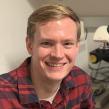
Dr. Walter focuses on splicing factor genes, which carry out the RNA splicing process and are widely mutated in lung cancer. The splicing factor U2AF1 is mutated in 2% of lung cancer patients, but 80% of these mutations are identical, making it one of the most common missense mutations in lung cancer. Scientists do not have a good understanding of why this mutation occurs, or how it promotes cancer development. Dr. Walter will use a combination of cell and mouse model systems along with patient data to identify the unique molecular and genetic features of U2AF1-mutant cancer cells with the goal of identifying new therapeutic targets for lung cancer patients.
Bo Gu, PhD
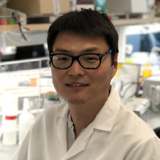
Dr. Gu [Fraternal Order of Eagles Fellow] is deciphering the combinatorial code of mammalian transcription regulation. The precise and robust regulation of gene expression is typically achieved through a combination of multiple transcription factors. However, we lack understanding of how a mammalian transcription system perceives, processes, and presents combinations of transcription factors. Dr. Gu will combine quantitative modeling and synthetic approaches to analyze the complex interactions among natural transcription regulatory proteins and apply the principles learned to engineer a programmable transcriptional platform with tunable logic. This work promises to deepen our understanding of mammalian transcription regulation and unlock new capabilities for emerging cell-based therapeutics.
Georgia R. Squyres, PhD
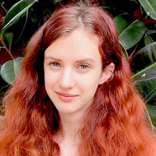
Dr. Squyres [National Mah Jongg League Fellow] is using quantitative microscopy and cell biology approaches to study how bacteria in biofilms coordinate their behavior in space and time. Biofilms are dense, multicellular communities of bacteria embedded in an extracellular matrix. Biofilms often form during bacterial infections, resulting in infections that are difficult to treat and resist antibiotics; cancer patients are at particular risk for these types of infections. Dr. Squyres is currently investigating how the release of extracellular DNA, a key component of the biofilm matrix, is coordinated during biofilm development. Greater understanding of how bacteria function in biofilms can lead to new approaches to target these treatment-resistant infections.
Catherine A. Freije, PhD
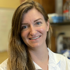
Dr. Freije [Berger Foundation Fellow] is studying how the genetic diversity of hepatitis B virus (HBV) is shaped by its need to replicate and interact with specific host genes. Current antiviral therapeutics for HBV merely suppress infection and do not cure disease; as a result, patients with chronic HBV infection are at risk of developing liver cancer. Dr. Freije plans to uncover essential genomic regions that HBV needs to survive and persist, as well as those that counteract host genes that function to restrict these activities. This approach could provide insight into the progression of disease and has the potential to identify new antiviral therapeutics and ultimately reduce the incidence of HBV-associated liver cancer.
Edie I. Crosse, PhD
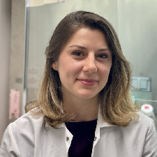
Myelodysplasia and acute myeloid leukemia are blood cancers with a poor prognosis. At the root of these malignancies are cells harboring mutant forms of proteins with dysfunctional activity which results in abnormal cell behavior and drives disease progression. The focus of my project is the development of new therapeutics that precisely identify cells with mutant forms of the proteins and, by harnessing their aberrant biological activity, causes those cells to self-destruct. These selective therapeutics will be able to kill cancer cells but leave the healthy cells intact proving more effective and having less side-effects than the chemotherapies currently in use.
Madi Y. Cissé, PhD

Dr. Cissé [Merck Fellow] aims to define the functional importance of nutrient sensing within the tumor microenvironment. How cells sense and adapt to the availability of nutrients in their environment is incompletely understood, but one key pathway is the signaling system anchored by the mTORC1 kinase. The mTORC1 kinase regulates cell growth and metabolism in response to nutrients such as amino acids and glucose. Aberrant mTORC1 signaling is implicated in several cancers, including melanoma, known to be heavily influenced by factors in the microenvironment such as nutrient availability. Dr. Cissé aims to understand how tumor metabolism senses and responds to varying nutrient levels, which will be essential for developing novel therapeutic targets.
Marco A. Catipovic, PhD
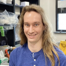
Dr. Catipovic [HHMI Fellow] focuses on the mechanisms governing the resolution of errors that arise during RNA translation in mammals. Ribosomes translating the same message can collide if they are damaged or encounter blockages much like cars involved in a traffic accident. While cells can tolerate small numbers of these incidents, pervasive collisions overwhelm the cell and force it to make crucial decisions regarding long-term viability. Dr. Catipovic investigates the biochemical mechanisms governing this determination. He uses reconstituted translation systems, consisting of purified translation factors in vitro, as a tool to study the signaling pathways initiated by ribosomal collisions that effect the life-death decisions of severely stressed cells. Perturbation of these pathways can cause premature cell death or unregulated cellular proliferation, which is found in almost all cancers.
